File list
From Slicer Wiki
This special page shows all uploaded files.
| Date | Name | Thumbnail | Size | Description | Versions |
|---|---|---|---|---|---|
| 21:39, 5 November 2013 | RSSLeftCaudateRejectionLabelNoLeakage.png (file) |  |
391 KB | 1 | |
| 21:38, 5 November 2013 | RSSLeftCaudateRejectionLabel.png (file) |  |
275 KB | 1 | |
| 21:37, 5 November 2013 | RSSLeftCaudateLeakage.png (file) |  |
405 KB | 1 | |
| 21:04, 5 November 2013 | MeningiomaRSSPanel.png (file) |  |
52 KB | 2 | |
| 20:55, 5 November 2013 | RSSPanelSlicer4.png (file) |  |
51 KB | 2 | |
| 20:44, 18 June 2013 | AfibSegmentationAidedRegistrationContour.png (file) |  |
570 KB | 1 | |
| 20:34, 18 June 2013 | AfibSegmentationAidedRegistration.png (file) |  |
566 KB | 1 | |
| 20:01, 18 June 2013 | AfibMIAffineRegistrationWithContour.png (file) |  |
696 KB | affine registration using MI metric of two LGE MRI. The red contour is the left atrium. We see that the overall registration is good, but the left atrium position is not accurate. In the case where we want to study the LA, this is not good enough. | 1 |
| 15:10, 18 June 2013 | AfibMIAffineRegistration.png (file) | 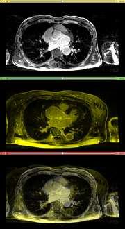 |
578 KB | Affine registration using MI metric of two MR images. | 1 |
| 15:05, 18 June 2013 | SegmentationAidedRegistrationUsageScreenShot.png (file) |  |
47 KB | 3 | |
| 13:46, 18 June 2013 | AfibBeforeRegistrationSmall.png (file) |  |
834 KB | two heart MRI images, axial, which are to be registered. Top: fixed image Middle: moving image Bottom: putting them together. | 1 |
| 23:41, 13 June 2013 | SegmentationAidedRegistrationComparison.png (file) | 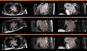 |
718 KB | 1 | |
| 02:58, 8 June 2013 | UAB MONOGRAM.png (file) |  |
5 KB | 1 | |
| 02:29, 8 June 2013 | PNLlogo4.png (file) |  |
703 KB | one logo of PNL | 1 |
| 15:11, 7 June 2013 | SegAidedRegSquareFocus128.png (file) |  |
25 KB | icon for SegmentationAidedRegistration extension | 1 |
| 15:54, 24 November 2011 | LUNGIXRSSSegmentation.png (file) |  |
758 KB | segment lung from CT image using RSS | 1 |
| 14:18, 24 November 2011 | KidneyLeftCTRSS.png (file) |  |
598 KB | segment left kidney from CT using RSS | 1 |
| 14:00, 24 November 2011 | KidneyRightCTRSS.png (file) |  |
519 KB | Extract right kidney from CT image using RSS | 1 |
| 11:54, 24 November 2011 | MeningiomaCase1RSSLabel.png (file) |  |
631 KB | the manual label map drawn for the meningioma case1 | 1 |
| 20:34, 23 November 2011 | MeningiomaCase1.png (file) |  |
622 KB | update with slicer4 gui | 3 |
| 14:30, 10 May 2011 | Rss CT liver.png (file) |  |
536 KB | 2 | |
| 14:23, 10 May 2011 | CT liver segmentation case.tgz (file) | 62.91 MB | 1 | ||
| 00:34, 28 October 2010 | Rss.pdf (file) |  |
572 KB | 1 | |
| 23:34, 26 April 2010 | RSSMandible.png (file) |  |
446 KB | 1 | |
| 14:45, 26 April 2010 | RSS-aorta.png (file) |  |
518 KB | segmentation of aorta using RSS | 1 |
| 20:15, 22 April 2010 | RssVentricle.png (file) |  |
761 KB | 2 | |
| 21:05, 14 April 2010 | RssPanel.png (file) | 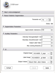 |
17 KB | 3 | |
| 20:50, 14 April 2010 | RSS run.png (file) |  |
490 KB | 1 | |
| 20:46, 14 April 2010 | RSS editStep.png (file) | 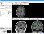 |
498 KB | 1 | |
| 20:35, 14 April 2010 | RSS tumor.png (file) |  |
579 KB | 1 | |
| 20:18, 14 April 2010 | RSS rkidney.png (file) |  |
736 KB | 1 | |
| 15:02, 14 April 2010 | RSSkidneyL.png (file) |  |
599 KB | 1 | |
| 00:34, 8 April 2010 | RobustStatisticsSegmentation usage3.png (file) | 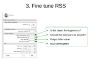 |
94 KB | 2 | |
| 00:33, 8 April 2010 | RobustStatisticsSegmentation usage2.png (file) |  |
86 KB | 2 | |
| 00:33, 8 April 2010 | RobustStatisticsSegmentation usage1.png (file) |  |
193 KB | 2 | |
| 03:56, 7 April 2010 | RSS MultiObjSeg1.png (file) |  |
240 KB | 1 |