Modules:JointRicianLMMSEImageFilter-Documentation-3.6
Return to Slicer 3.6 Documentation
Module Name
jointLMMSE
General Information
Module Type & Category
Type: CLI
Category: Diffusion MRI Applications
Authors, Collaborators & Contact
- Author: Antonio Tristán Vega and Santiago Aja Fernández
- Contact: atriveg@bwh.harvard.edu
Module Description
This module reduces Rician noise (or unwanted detail) on a set of diffusion weighted images. For this, it filters the image in the mean squared error sense using a Rician noise model. The N closest gradient directions to the direction being processed are filtered together to improve the results: the noise-free signal is seen as an n-diemensional vector which has to be estimated with the LMMSE method from a set of corrupted measurements. To that end, the covariance matrix of the noise-free vector and the cross covariance between this signal and the noise have to be estimated, which is done taking into account the image formation process.
The noise parameter is automatically estimated from a rough segmentation of the background of the image. In this area the signal is simply 0, so that Rician statistics reduce to Rayleigh and the noise power can be easily estimated from the mode of the histogram.
A complete description of the algorithm may be found in:
Antonio Tristán-Vega and Santiago Aja-Fernández, "DWI filtering using joint information for DTI and HARDI", Medical Image Analysis, Volume 14, Issue 2, Pages 205-218. 2010.
Usage
The filter operates on an input DWI volume (gradient directions must be known). The meaning of the parameters of this module is listed below.
Examples, Use Cases & Tutorials
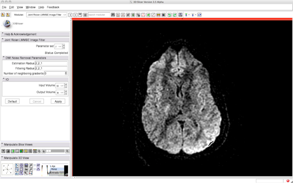
The following screenshots illustrate how to use the filter and the expected results (note that the optimal parameters strongly depends on the characteristics of the image: voxel size, number of gradients...).
Before filtering:
After filtering:
Quick Tour of Features and Use
It is very easy to use it. Just select a DWI, set the parameters (if you really need it), and you're ready to go.
- Input DWI Volume: set the DWI volume
- Output DWI Volume: the filtered DWI volume
- Estimation radius: This is the 3D radius of the neighborhood used for noise estimation. Noise power is estimated as the mode of the histogram of local variances
- Filtering radius: This is the 3D radius of the neighborhood used for filtering: local means and covariance matrices are estimated within this neighborhood. A large radius more effectively reduces the noise but may induce a certain blurring of the edges.
- Number of neighborhood gradients: This filter works gathering joint information from the N closest gradient directions to the one under study. This parameter is N. If N=0 is fixed, then all gradient directions are filtered together.
- NOTE: If N=1 is used this filter is similar (but not equal) to RicianLMMSEImageFilter. Two main differences exist: 1) 4-th order moments have to be computed only for baseline(s) image(s), and 2) if more than one baseline is present all of them are filtered together even if N=1.
Development
Dependencies
Volumes. Needed to load DWI volumes
Known bugs
Usability issues
Source code & documentation
Source Code: Follow this link
Doxygen documentation:
More Information
Acknowledgment
Antonio Tristan Vega, Santiago Aja Fernandez. University of Valladolid (SPAIN). Partially funded by grant number TEC2007-67073/TCM from the Ministerio de Ciencia e Innovación (Spain) and "FEDER" European Regional Development Fund.
References
- Antonio Tristán-Vega and Santiago Aja-Fernández, "DWI filtering using joint information for DTI and HARDI", Medical Image Analysis, Volume 14, Issue 2, Pages 205-218 (2010).
- Santiago Aja-Fernández, Antonio Tristán-Vega, and Carlos Alberola-López, "Noise estimation in single- and multiple-coil magnetic resonance data based on statistical models". Magnetic Resonance Imaging, Volume 27, Pages 1397-1409 (2009).

