Main Page/SlicerCommunity/2018
Go to 2022 :: 2021 :: 2020 :: 2019 :: 2018 :: 2017 :: 2016 :: 2015 :: 2014-2011 :: 2010-2000
The community that relies on 3D Slicer is large and active: (numbers below updated on December 1st, 2023)
- 1,467,466+ downloads in the last 11 years (269,677 in 2023, 206,541 in 2022)
- over 17.900+ literature search results on Google Scholar
- 2,147+ papers on PubMed citing the Slicer platform paper
- Fedorov A., Beichel R., Kalpathy-Cramer J., Finet J., Fillion-Robin J-C., Pujol S., Bauer C., Jennings D., Fennessy F.M., Sonka M., Buatti J., Aylward S.R., Miller J.V., Pieper S., Kikinis R. 3D Slicer as an Image Computing Platform for the Quantitative Imaging Network. Magnetic Resonance Imaging. 2012 Nov;30(9):1323-41. PMID: 22770690. PMCID: PMC3466397.
- 39 events in open source hackathon series continuously running since 2005 with 3260 total participants
- Slicer Forum with +8,138 subscribers has approximately 275 posts every week
The following is a sample of the research performed using 3D Slicer outside of the group that develops it. in 2018
We monitor PubMed and related databases to update these lists, but if you know of other research related to the Slicer community that should be included here please email: marianna (at) bwh.harvard.edu.
Contents
- 1 2018
- 1.1 From 3D Imaging to 3D Printing in Dentistry - A Practical Guide
- 1.2 Gray Matter Atrophy in Multiple Sclerosis Despite Clinical and Lesion Stability During Natalizumab Treatment
- 1.3 Thalamic Shape and Volume Abnormalities in Female Patients with Panic Disorder
- 1.4 Axillary Lymph Node Evaluation Utilizing Convolutional Neural Networks Using MRI Dataset
- 1.5 Dosimetric Evaluation of Respiratory Gated Volumetric Modulated Arc Therapy for Lung Stereotactic Body Radiation Therapy using 3D Printing Technology
- 1.6 Pre-Stroke Surgery is not Beneficial to Normotensive Rats Undergoing Sixty Minutes of Transient Focal Cerebral Ischemia
- 1.7 Patient-specific Interactive Software Module for Virtual Preoperative Planning and Visualization of Pedicle Screw Entry Point and Trajectories in Spine Surgery
- 1.8 Detectability of Radiation-induced Changes in Magnetic Resonance Biomarkers following Stereotactic Radiosurgery: A Pilot Study
- 1.9 Smaller Volumes in the Lateral and Basal Nuclei of the Amygdala in Patients with Panic Disorder
- 1.10 A Novel Tool for Supervised Segmentation Using 3D Slicer
- 1.11 Real-time, Image-based Slice-to-volume Registration for Ultrasound-guided Spinal Intervention
- 1.12 Computed Tomographic Evaluation of Cleft Palate in One-day-old Puppies
- 1.13 A Wearable Mixed-reality Holographic Computer for Guiding External Ventricular Drain Insertion at the Bedside
- 1.14 The Dark Shades of the Antikythera Mechanism
- 1.15 Lung Tumor Segmentation Methods: Impact on the Uncertainty of Radiomics Features for Non-small Cell Lung Cancer
- 1.16 Accuracy Assessment of 3D-Printed Tooth Replicas
- 1.17 Non-invasive Magnetic Resonance Imaging of Oils in Botryococcus Braunii Green Algae: Chemical Shift Selective and Diffusion-weighted Imaging
- 1.18 Altered White Matter Connectivity in Patients with Schizophrenia: An Investigation using Public Neuroimaging Data from SchizConnect
- 1.19 SlicerSALT: Shape AnaLysis Toolbox
- 1.20 Association of Anemia and Hemoglobin Decrease during Acute Stroke Treatment with Infarct Growth and Clinical Outcome
- 1.21 Outer Wall Segmentation of Abdominal Aortic Aneurysm by Variable Neighborhood Search Through Intensity and Gradient Spaces
- 1.22 Simple and Robust Referencing System Enables Identification of Dissolved-phase Xenon Spectral Frequencies
- 1.23 Diffusion Weighted And Dynamic Contrast Enhanced MRI as an Imaging Biomarker for Stereotactic Ablative Body Radiotherapy (SABR) of Primary Renal Cell Carcinoma
- 1.24 Toward a Real-time System for Temporal Enhanced Ultrasound-guided Prostate Biopsy
- 1.25 Innovations in Preoperative Planning: Insights into Another Dimension using 3D Printing for Cardiac Disease
- 1.26 3D-constructive Interference into Steady State (3D-CISS) Labyrinth Signal Alteration in Patients with Vestibular Schwannoma
- 1.27 Computed Tomography Trachea Volumetry in Patients With Scleroderma: Association With Clinical and Functional Findings
- 1.28 Comparison of Modified Two-point Dixon and Chemical Shift Encoded MRI Water-fat Separation Methods for Fetal Fat Quantification
- 1.29 Genetic Mapping of Molar Size Relations Identifies Inhibitory Locus for Third Molars in Mice
- 1.30 Characterization of Adrenal Lesions on Unenhanced MRI Using Texture Analysis: A Machine-Learning Approach
- 1.31 Role of CT and MRI in the Design and Development of Orthopaedic Model using Additive Manufacturing
- 1.32 Comparing Damage on Retrieved Total Elbow Replacement Bushings with Lab Worn Specimens Subjected to Varied Loading Conditions
- 1.33 Validation of a Windowing Protocol for Accurate In Vivo Tooth Segmentation Using i-Cat Cone Beam Computed Tomography
- 1.34 Automatic Segmentation of Stereoelectroencephalography (SEEG) Electrodes Post-Implantation Considering Bending
- 1.35 Low-Cost Three-Dimensional Printed Phantom for Neuraxial Anesthesia Training: Development and Comparison to a Commercial Model
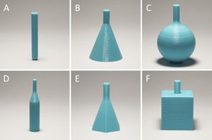
- 1.36 Improvement of Quality of 3D Printed Objects by Elimination of Microscopic Structural Defects in Fused Deposition Modeling
- 1.37 Targeting HER2 Aberrations in Non-Small Cell Lung Cancer with Osimertinib
- 1.38 Plasma Membrane LAT Activation Precedes Vesicular Recruitment Defining Two Phases of Early T-cell Activation
- 1.39 Advances in Stereotactic Navigation for Pelvic Surgery
- 1.40 New Approach of Ultra-focal Brachytherapy for Low- and Intermediate-risk Prostate Cancer with Custom-linked I-125 Seeds: A feasibility Study of Optimal Dose Coverage
- 1.41 Evaluating the Association between Enlarged Perivascular Spaces and Disease Worsening in Multiple Sclerosis
- 1.42 Clinical Evaluation of Semi-Automatic Open-Source Algorithmic Software Segmentation of the Mandibular Bone: Practical Feasibility and Assessment of a New Course of Action
- 1.43 Subthalamic Oscillatory Activity and Connectivity during Gait in Parkinson's Disease
- 1.44 Complete Thoracolumbar Fracture-dislocation with Intact Neurologic Function: Explanation of a Novel Cord Saving Mechanism
- 1.45 Optimization of 3D Print Material for the Recreation of Patient-Specific Temporal Bone Models
- 1.46 3-D Segmentation of Lung Nodules using Hybrid Level Sets
- 1.47 Prediction of Outcome using Pretreatment 18F-FDG PET/CT and MRI Radiomics in Locally Advanced Cervical Cancer Treated with Chemoradiotherapy
- 1.48 Smoking Duration Alone Provides Stronger Risk Estimates of Chronic Obstructive Pulmonary Disease than Pack-years
- 1.49 Using 3DSlicer, Z-Brush, and Slic3r to Turn CAT Scans Into Kidney 3D-Prints
- 1.50 Development of White Matter Microstructure in Relation to Verbal and Visuospatial Working Memory-A Longitudinal Study
- 1.51 Detailed T1-Weighted Profiles from the Human Cortex Measured in Vivo at 3 Tesla MRI
- 1.52 Estimating Shape Correspondence for Populations of Objects with Complex Topology
- 1.53 Development of a Hybrid Computational/Experimental Framework for Evaluation of Damage Mechanisms of a Linked Semiconstrained Total Elbow System
- 1.54 Simulation of the Human Airways using Virtual Topology Tools and Meshing Optimization
- 1.55 Planning of Skull Reconstruction Based on a Statistical Shape Model Combined with Geometric Morphometrics
- 1.56 Contribution of 3D Printing to Mandibular Reconstruction after Cancer
- 1.57 Optimizing Image Quantification for 177Lu SPECT/CT Based on a 3D Printed 2-Compartment Kidney Phantom
- 1.58 Endocardial Infarct Scar Recognition by Myocardial Electrical Impedance is not Influenced by Changes in Cardiac Activation Sequence
- 1.59 Regional Hippocampal Vulnerability in Early Multiple Sclerosis: Dynamic Pathological Spreading from Dentate Gyrus to CA1
- 1.60 Effects of Total Saponins from Trillium Tschonoskii Rhizome on Grey and White Matter Injury Evaluated by Quantitative Multiparametric MRI in a Rat Model of Ischemic Stroke
- 1.61 Optimized Programming Algorithm for Cylindrical and Directional Deep Brain Stimulation Electrodes
- 1.62 Temporomandibular Joint Regeneration: Proposal of a Novel Treatment for Condylar Resorption after Orthognathic Surgery using Transplantation of Autologous Nasal Septum Chondrocytes, and the First Human Case Report
- 1.63 Validation of MRI to TRUS Registration for High-dose-rate Prostate Brachytherapy
- 1.64 Commissioning and Validation of Commercial Deformable Image Registration Software for Adaptive Contouring
- 1.65 Performance of Ultrafast DCE-MRI for Diagnosis of Prostate Cancer
- 1.66 A Cost-Effective, In-House, Positioning and Cutting Guide System for Orthognathic Surgery
- 1.67 Comparison of 3D Echocardiogram-Derived 3D Printed Valve Models to Molded Models for Simulated Repair of Pediatric Atrioventricular Valves
- 1.68 A Preliminary Study on Precision Image Guidance for Electrode Placement in an EEG Study
- 1.69 Radiologic Factors Predicting Deterioration of Mental Status in Patients with Acute Traumatic Subdural Hematoma
- 1.70 How to Precisely Measure the Volume Velocity Transfer Function of Physical Vocal Tract Models by External Excitation
- 1.71 Longitudinal Microstructural Changes of Cerebral White Matter and their Association with Mobility Performance in Older Persons
- 1.72 Micro-computed Tomographic Evaluation of the Shaping Ability of XP-endo Shaper, iRaCe, and EdgeFile Systems in Long Oval-shaped Canals
- 1.73 Cerebral Radiation Necrosis: An Analysis of Clinical and Quantitative Imaging and Volumetric Features
- 1.74 Nonlinear Deformation of Tractography in Ultrasound-guided Low-grade Gliomas Resection
- 1.75 Lateral Ventricular Volume Asymmetry Predicts Poor Outcome After Spontaneous Intracerebral Hemorrhage
- 1.76 High Fidelity Virtual Reality Orthognathic Surgery Simulator
- 1.77 Volumetric Analysis of Magnetic Resonance-guided Focused Ultrasound Thalamotomy Lesions
- 1.78 SVA: Shape Variation Analyzer
- 1.79 Automatic Quantification Framework to Detect Cracks in Teeth
- 1.80 Detection of Bone Loss via Subchondral Bone Analysis
- 1.81 Development of a Patient-specific Tumor Mold using Magnetic Resonance Imaging and 3-Dimensional Printing Technology for Targeted Tissue Procurement and Radiomics Analysis of Renal Masses
- 1.82 A Surface-based Approach to Determine Key Spatial Parameters of the Acetabulum in a Standardized Pelvic Coordinate System
- 1.83 Imaging of Concussion in Young Athletes
- 1.84 Advantages and Disadvantages in Image Processing with Free Software in Radiology
- 1.85 WRIST: A WRist Image Segmentation Toolkit for Carpal bone Delineation from MRI
- 1.86 Enhanced Preoperative Deep Inferior Epigastric Artery Perforator Flap Planning with a 3D-Printed Perforasome Template: Technique and Case Report
- 1.87 DCE-MRI Pharmacokinetic-Based Phenotyping of Invasive Ductal Carcinoma: A Radiomic Study for Prediction of Histological Outcomes
- 1.88 MRI-Based Experimentations of Fingertip Flat Compression: Geometrical Measurements and Finite Element Inverse Simulations to Investigate Material Property Parameters
- 1.89 Objective Assessment of Colonoscope Manipulation Skills in Colonoscopy Training
- 1.90 Primary Trigeminal Neuralgia is Associated with Posterior Fossa Crowdedness: A Prospective Case-Control Study
- 1.91 Bone Marrow Drives Central Nervous System Regeneration after Radiation Injury
- 1.92 Quantitative Spinal Cord MRI in Radiologically Isolated Syndrome
- 1.93 Radiogenomic Analysis of Hypoxia Pathway is Predictive of Overall Survival in Glioblastoma
- 1.94 Paracrine Osteoprotegerin and β-catenin Stabilization Support Synovial Sarcomagenesis in Periosteal Cells
- 1.95 A New Device for Fiducial Registration of Image-guided Navigation System for Liver RFA
- 1.96 Uncoupling N-acetylaspartate from Brain pathology: Implications for Canavan Disease Gene Therapy
2018
From 3D Imaging to 3D Printing in Dentistry - A Practical Guide
|
Publication: Int J Comput Dent. 2018;21(4):345-56. PMID: 30539177. Authors: Moser N, Santander P, Quast A. Institution: Department of Oral and Maxillofacial Surgery, University Medicine Göttingen, Göttingen, Germany. Abstract: 3D imaging in dentistry plays an essential part in diagnostics and treatment planning. To transform digital images into a real object that can be experienced haptically may provide new opportunities to practitioners regarding patient communication, skills training, and treatment planning. Therefore, the aim of this article is to provide a practical guide from 3D imaging to 3D printing using low-cost printers and open source software; the authors used 3D Slicer software and a Meshmixer printer, including the printer's own software. The article presents step-by-step instructions on how to perform rapid prototyping via fused deposition modeling (FDM) and stereolithography (SLA). As an example, we printed the skull of a patient with Saethre-Chotzen syndrome who was undergoing maxillofacial surgery. The protocol explained here should enable the technically interested clinician to produce patient-specific 3D models in-house, prefabricate osteosynthesis plates, and take advantage of the benefits of 3D printing for dentist-patient communication. |
Gray Matter Atrophy in Multiple Sclerosis Despite Clinical and Lesion Stability During Natalizumab Treatment
|
Publication: PLoS One. 2018 Dec 21;13(12):e0209326. PMID: 30576361 | PDF Authors: Koskimäki F, Bernard J, Yong J, Arndt N, Carroll T, Lee SK, Reder AT, Javed A. Institution: Division of Clinical Neurosciences, Turku University Hospital and University of Turku, Turku, Finland. Abstract: Brain volume loss is an important surrogate marker for assessing disability in MS; however, contribution of gray and white matter to the whole brain volume loss needs further examination in the context of specific MS treatment. OBJECTIVES: To examine whole and segmented gray, white, thalamic, and corpus callosum volume loss in stable patients receiving natalizumab for 2-5 years. METHODS: This was a retrospective study of 20 patients undergoing treatment with natalizumab for 24-68 months. Whole brain volume loss was determined with SIENA. Gray and white matter segmentation was done using FAST. Thalamic and corpus callosum volumes were determined using Freesurfer. T1 relaxation values of chronic hypointense lesions (black holes) were determined using a quantitative, in-house developed method to assess lesion evolution. "...FLAIR images were used to determine T2 lesion volume using 3D Slicer. RESULTS: Over a mean of 36.6 months, median percent brain volume change (PBVC) was -2.0% (IQR 0.99-2.99). There was decline in gray (p = 0.001) but not white matter (p = 0.6), and thalamic (p = 0.01) but not corpus callosum volume (p = 0.09). Gray matter loss correlated with PBVC (Spearman's r = 0.64, p = 0.003) but not white matter (Spearman's r = 0.42, p = 0.07). Age significantly influenced whole brain volume loss (p = 0.010, multivariate regression), but disease duration and baseline T2 lesion volume did not. There was no change in T1 relaxation values of lesions or T2 lesion volume over time. All patients remained clinically stable. CONCLUSIONS: These results demonstrate that brain volume loss in MS is primarily driven by gray matter changes and may be independent of clinically effective treatment. |
Thalamic Shape and Volume Abnormalities in Female Patients with Panic Disorder
|
Publication: PLoS One. 2018 Dec 19;13(12):e0208152. PMID: 30566534 | PDF Authors: Asami T, Yoshida H, Takaishi M, Nakamura R, Yoshimi A, Whitford TJ, Hirayasu Y. Institution: Department of Psychiatry, Graduate School of Medicine, Yokohama City University, Yokohama, Japan. Abstract: The thalamus is believed to play crucial role in processing viscero-sensory information, and regulating the activity of amygdala in patients with panic disorder (PD). Previous functional neuroimaging studies have detected abnormal activation in the thalamus in patients with PD compared with healthy control subjects (HC). Very few studies, however, have investigated for volumetric abnormalities in the thalamus in patients with PD. Furthermore, to the best of our knowledge, no previous study has investigated for shape abnormalities in the thalamus in patients with PD. Twenty-five patients with PD and 25 HC participants (all female) were recruited for the study. A voxel-wise volume comparison analysis and a vertex-wise shape analysis were conducted to evaluate structural abnormalities in the PD patients compared to HC. The patients with PD demonstrated significant gray matter volume reductions in the thalamus bilaterally, relative to the HC. The shape analysis detected significant inward deformation in some thalamic regions in the PD patients, including the anterior nucleus, mediodorsal nucleus, and pulvinar nucleus. PD patients showed shape deformations in key thalamic regions that are believed to play a role in regulating emotional and cognitive functions. Funding:
|
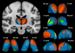 Female patients with panic disorder showed significant inward deformation of shape in the bilateral thalamus compared with female healthy control subjects (false discovery rate corrected, P < .05). These regions included (in the right thalamus) the anterior nucleus, medial mediodorsal nucleus, and lateral posterior nuclei (orange), and the medial part of pulvinar nucleus (green). In the left thalamus, the anterior nucleus, ventro-lateral nucleus, ventral anterior nucleus (orange), the medial part of pulvinar nucleus (green), and the lateral part of pulvinar nucleus (yellow) were affected. Abbreviations: Rt, right; Lt, left; ant, anterior view; post, posterior view; lat, lateral view; med, medial view; sup, superior view; inf, inferior view; 3D images were created using 3D Slicer. |
Axillary Lymph Node Evaluation Utilizing Convolutional Neural Networks Using MRI Dataset
|
Publication: J Digit Imaging. 2018 Dec;31(6):851-6. PMID: 29696472 | PDF Authors: Ha R, Chang P, Karcich J, Mutasa S, Fardanesh R, Wynn RT, Liu MZ, Jambawalikar S. Institution: Department of Radiology, Columbia University Medical Center, New York, NY, USA. Abstract: The aim of this study is to evaluate the role of convolutional neural network (CNN) in predicting axillary lymph node metastasis, using a breast MRI dataset. An institutional review board (IRB)-approved retrospective review of our database from 1/2013 to 6/2016 identified 275 axillary lymph nodes for this study. Biopsy-proven 133 metastatic axillary lymph nodes and 142 negative control lymph nodes were identified based on benign biopsies (100) and from healthy MRI screening patients (42) with at least 3 years of negative follow-up. For each breast MRI, axillary lymph node was identified on first T1 post contrast dynamic images and underwent 3D segmentation using an open source software platform 3D Slicer. A 32 × 32 patch was then extracted from the center slice of the segmented tumor data. A CNN was designed for lymph node prediction based on each of these cropped images. The CNN consisted of seven convolutional layers and max-pooling layers with 50% dropout applied in the linear layer. In addition, data augmentation and L2 regularization were performed to limit overfitting. Training was implemented using the Adam optimizer, an algorithm for first-order gradient-based optimization of stochastic objective functions, based on adaptive estimates of lower-order moments. Code for this study was written in Python using the TensorFlow module (1.0.0). Experiments and CNN training were done on a Linux workstation with NVIDIA GTX 1070 Pascal GPU. Two class axillary lymph node metastasis prediction models were evaluated. For each lymph node, a final softmax score threshold of 0.5 was used for classification. Based on this, CNN achieved a mean five-fold cross-validation accuracy of 84.3%. It is feasible for current deep CNN architectures to be trained to predict likelihood of axillary lymph node metastasis. Larger dataset will likely improve our prediction model and can potentially be a non-invasive alternative to core needle biopsy and even sentinel lymph node evaluation. |
Dosimetric Evaluation of Respiratory Gated Volumetric Modulated Arc Therapy for Lung Stereotactic Body Radiation Therapy using 3D Printing Technology
|
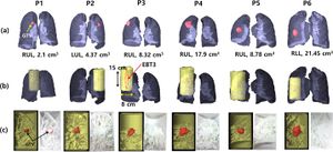
Publication: PLoS One. 2018 Dec 26;13(12):e0208685. PMID: 30586367 | PDF Authors: Yoon K, Jeong C, Kim SW, Cho B, Kwak J, Kim SS, Song SY, Choi EK, Ahn S, Lee SW. Institution: Department of Radiation Oncology, Asan Medical Center, University of Ulsan College of Medicine, Seoul, Republic of Korea. Abstract: PURPOSE: This study aimed to evaluate the dosimetric accuracy of respiratory gated volumetric modulated arc therapy (VMAT) for lung stereotactic body radiation therapy (SBRT) under simulation conditions similar to the actual clinical situation using patient-specific lung phantoms and realistic target movements. METHODS: Six heterogeneous lung phantoms were fabricated using a 3D-printer (3DISON, ROKIT, Seoul, Korea) to be dosimetrically equivalent to actual target regions of lung SBRT cases treated via gated VMAT. They were designed to move realistically via a motion device (QUASAR, Modus Medical Devices, Canada). Using the lung phantoms and a homogeneous phantom (model 500-3315, Modus Medical Devices), film dosimetry was performed with and without respiratory gating for VMAT delivery (TrueBeam STx; Varian Medical Systems, Palo Alto, CA, USA). The measured results were analyzed with the gamma passing rates (GPRs) of 2%/1 mm criteria, by comparing with the calculated dose via the AXB and AAA algorithms of the Eclipse Treatment Planning System (version 10.0.28; Varian Medical Systems). RESULTS: GPRs were greater than the acceptance criteria 80% for all film measurements with the stationary and homogeneous phantoms in conventional QAs. Regardless of the heterogeneity of phantoms, there were no significant differences (p > 0.05) in GPRs obtained with and without target motions; the statistical significance (p = 0.031) was presented between both algorithms under the utilization of heterogeneous phantoms. CONCLUSIONS: Dosimetric verification with heterogeneous patient-specific lung phantoms could be successfully implemented as the evaluation method for gated VMAT delivery. In addition, it could be dosimetrically confirmed that the AXB algorithm improved the dose calculation accuracy under patient-specific simulations using 3D printed lung phantoms. "The DICOM files including segmented contours were imported to the 3D Slicer program , in which the geometric information of contours was converted to a Standard Tessellation Language (STL) format." Funding:
|
Pre-Stroke Surgery is not Beneficial to Normotensive Rats Undergoing Sixty Minutes of Transient Focal Cerebral Ischemia
|
Publication: PLoS One. 2018 Dec 28;13(12):e0209370. PMID: 30592760 | PDF Authors: Bayliss M, Trotman-Lucas M, Janus J, Kelly ME, Gibson CL. Institution: Department of Neuroscience, Psychology & Behaviour, University of Leicester, Leicester, United Kingdom. Abstract: Experimental stroke in rodents, via middle cerebral artery occlusion (MCAO), can be associated with a negative impact on wellbeing and mortality. In hypertensive rodents, pre-stroke craniotomy increased survival and decreased body weight loss post-MCAO. Here we determined the effect, in normotensive Sprague-Dawley rats following 60 minutes MCAO, with or without pre-surgical craniotomy, on post-stroke outcomes in terms of weight loss, neurological deficit, lesion volume and functional outcomes. There was no effect of pre-stroke craniotomy on indicators of wellbeing including survival rate (P = 0.32), body weight loss (P = 0.42) and neurological deficit (P = 0.75). We also assessed common outcome measures following experimental stroke and found no effect of pre-stroke craniotomy on lesion volume as measured by T2-weighted MRI (P = 0.846), or functional performance up to 28 days post-MCAO (staircase test, P = 0.32; adhesive sticker test, P = 0.49; cylinder test, P = 0.38). Thus, pre-stroke craniotomy did not improve animal welfare in terms of body weight loss and neurological deficit. However, it is important, given that a number of drug delivery studies utilise the craniotomy procedure, to note that there was no effect on lesion volume or functional outcome following experimental stroke. "All images were corrected for intensity inhomogeneities introduced by the 2-channel surface receive coil using the bias field correction method in 3D Slicer, v.3.6." Funding:
|
Patient-specific Interactive Software Module for Virtual Preoperative Planning and Visualization of Pedicle Screw Entry Point and Trajectories in Spine Surgery
|
Publication: Neurol India. 2018 Nov-Dec;66(6):1766-70. PMID: 30504578 | PDF Authors: Muralidharan V, Swaminathan G, Devadhas D, Joseph BV. Institution: Department of Neurological Sciences, Christian Medical College, Vellore, Tamil Nadu, India. Abstract: BACKGROUND: Lumbar pedicle screw insertion involves a steep learning curve for novice spine surgeons and requires image guidance or navigation. Small volume centers may be handicapped by the lack of cost-effective user-friendly tools for preoperative planning, guidance, and decision making. OBJECTIVE: We describe a patient-specific interactive software module, pedicle screw simulator (PSS), for virtual preoperative planning to determine the entry point and visualize the trajectories of pedicle screws. MATERIALS AND METHODS: The PSS was coded in Python for use in an open source image processing software, 3D Slicer. Preoperative computed tomography (CT) data of each subject was loaded into this module. The entry-target (ET) mode calculates the ideal angle from the entry point through the widest section of the pedicle to the desired target in the vertebral body. The entry-angle (EA) mode projects the screw trajectory from the desired entry point at a desired angle. The performance of this software was tested using CT data from four subjects. RESULTS: PSS provided a quantitative and qualitative feedback preoperatively to the surgeon about the entry point and trajectories of pedicle screws. It also enabled the surgeons to visualize and predict the pedicle breach with various trajectories. CONCLUSION: This interactive software module aids in understanding and correcting the orientation of each vertebra in three-dimensions, to identify the ideal entry points, angles of insertion and trajectories for pedicle screw insertion to suit the local anatomy. |
 EA mode of pedicle screw trajectory planning in a patient with high grade L5-S1 spondylolisthesis. (a) Correction of vertebra rotation. (b) Marking of fiducials. (c) Desired trajectory for selected entry point and angle. (d) Marking of the entry and target fiducials for desired iliac wing screw. (e) Ideal trajectory of iliac wing screw and panel displaying the angle of screw trajectory using entry-target mode. |
Detectability of Radiation-induced Changes in Magnetic Resonance Biomarkers following Stereotactic Radiosurgery: A Pilot Study
|
Publication: PLoS One. 2018 Nov 26;13(11):e0207933. PMID: 30475887 | PDF Authors: Winter JD, Moraes FY, Chung C, Coolens C. Institution: Radiation Medicine Program, Princess Margaret Cancer Center and University Health Network, Toronto, Ontario, Canada. Abstract: Our objective was to investigate direct voxel-wise relationship between dose and early MR biomarker changes both within and in the high-dose region surrounding brain metastases following stereotactic radiosurgery (SRS). Specifically, we examined the apparent diffusion coefficient (ADC) from diffusion-weighted imaging and the contrast transfer coefficient (Ktrans) and volume of extracellular extravascular space (ve) derived from dynamic contrast-enhanced (DCE) MRI data. We investigated 29 brain metastases in 18 patients using 3 T MRI to collect imaging data at day 0, day 3 and day 20 following SRS. The ADC maps were generated by the scanner and Ktrans and ve maps were generated using in-house software for dynamic tracer-kinetic analysis. To enable spatially-correlated voxel-wise analysis, we developed a registration pipeline to register all ADC, Ktrans and ve maps to the planning MRI scan. To interrogate longitudinal changes, we computed absolute ΔADC, ΔKtrans and Δve for day 3 and 20 post-SRS relative to day 0. We performed a Kruskall-Wallice test on each biomarker between time points and investigated dose correlations within the gross tumour volume (GTV) and surrounding high dose region > 12 Gy via Spearman's rho. Only ve exhibited significant differences between day 0 and 20 (p < 0.005) and day 3 and 20 (p < 0.05) within the GTV following SRS. Strongest dose correlations were observed for ADC within the GTV (rho = 0.17 to 0.20) and weak correlations were observed for ADC and Ktrans in the surrounding > 12 Gy region. Both ΔKtrans and Δve showed a trend with dose at day 20 within the GTV and > 12 Gy region (rho = -0.04 to -0.16). Weak dose-related decreases in Ktrans and ve within the GTV and high dose region at day 20 most likely reflect underlying vascular responses to radiation. Our study also provides a voxel-wise analysis schema for future MR biomarker studies with the goal of elucidating surrogates for radionecrosis. "To enable spatially registered voxel-wise analyses, we developed a rigorous in-house registration pipeline to perform all image registration steps as well as visualize image registration results using Python (Python Software Foundation), interacting with 3D Slicer." Funding:
|
Smaller Volumes in the Lateral and Basal Nuclei of the Amygdala in Patients with Panic Disorder
|
Publication: PLoS One. 2018 Nov 7;13(11):e0207163. PMID: 30403747 | PDF Authors: Asami T, Nakamura R, Takaishi M, Yoshida H, Yoshimi A, Whitford TJ, Hirayasu Y. Institution: Department of Psychiatry, Graduate School of Medicine, Yokohama City University, Yokohama, Japan. Abstract: The amygdala plays an important functional role in fear and anxiety. Abnormalities in the amygdala are believed to be involved in the neurobiological basis of panic disorder (PD). Previous structural neuroimaging studies have found global volumetric and morphological abnormalities in the amygdala in patients with PD. Very few studies, however, have explored for structural abnormalities in various amygdala sub-regions, which consist of various sub-nuclei, each with different functions. This study aimed to evaluate for volumetric abnormalities in the amygdala sub-nuclei, in order to provide a better understanding neurobiological basis of PD. Thirty-eight patients with PD and 38 matched healthy control (HC) participants underwent structural MRI scanning. The volume of the whole amygdala, as well as its consistent sub-nuclei, were calculated using FreeSurfer software. Relative volumes of these amygdala sub-regions were compared between the two groups. Results showed significantly smaller volumes in the right lateral and basal nuclei in the patients with PD compared with the HC. Lateral and basal nuclei are thought to play crucial role for processing sensory information related with anxiety and fear. Our results suggest that these particular amygdala sub-regions play a role in the development of PD symptoms. Funding:
|
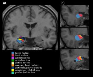 Nuclei of the amygdala. a) 3D image was constructed using 3D Slicer. b) Coronal images of the nuclei in the right amygdala. |
A Novel Tool for Supervised Segmentation Using 3D Slicer
|
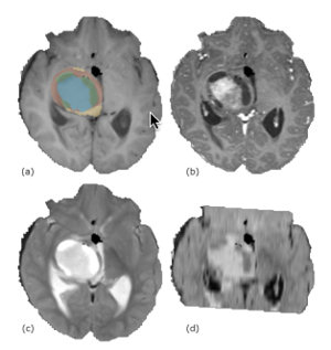
Publication: Symmetry 2018 Nov 12; 10:627. | PDF Authors: Daniel Chalupa, Jan Mikulka. Institution: Department of Theoretical and Experimental Electrical Engineering, Brno University of Technology, Brno, Czech Republic. Abstract: The rather impressive extension library of medical image-processing platform 3D Slicer lacks a wide range of machine-learning toolboxes. The authors have developed such a toolbox that incorporates commonly used machine-learning libraries. The extension uses a simple graphical user interface that allows the user to preprocess data, train a classifier, and use that classifier in common medical image-classification tasks, such as tumor staging or various anatomical segmentations without a deeper knowledge of the inner workings of the classifiers. A series of experiments were carried out to showcase the capabilities of the extension and quantify the symmetry between the physical characteristics of pathological tissues and the parameters of a classifying model. These experiments also include an analysis of the impact of training vector size and feature selection on the sensitivity and specificity of all included classifiers. The results indicate that training vector size can be minimized for all classifiers. Using the data from the Brain Tumor Segmentation Challenge, Random Forest appears to have the widest range of parameters that produce sufficiently accurate segmentations, while optimal Support Vector Machines’ training parameters are concentrated in a narrow feature space.. Funding:
|
Real-time, Image-based Slice-to-volume Registration for Ultrasound-guided Spinal Intervention
|
Publication: Phys Med Biol. 2018 Oct 29;63(21):215016. PMID: 30372418 | PDF Authors: De Silva T, Uneri A, Zhang X, Ketcha M, Han R, Sheth N, Martin A, Vogt S, Kleinszig G, Belzberg A, Sciubba DM, Siewerdsen JH. Institution: Department of Biomedical Engineering, Johns Hopkins University, Baltimore, MD, USA. Abstract: Real-time fusion of magnetic resonance (MR) and ultrasound (US) images could facilitate safe and accurate needle placement in spinal interventions. We develop an entirely image-based registration method (independent of or complementary to surgical trackers) that includes an efficient US probe pose initialization algorithm. The registration enables the simultaneous display of 2D ultrasound image slices relative to 3D pre-procedure MR images for navigation. A dictionary-based 3D-2D pose initialization algorithm was developed in which likely probe positions are predefined in a dictionary with feature encoding by Haar wavelet filters. Feature vectors representing the 2D US image are computed by scaling and translating multiple Haar basis filters to capture scale, location, and relative intensity patterns of distinct anatomical features. Following pose initialization, fast 3D-2D registration was performed by optimizing normalized cross-correlation between intra- and pre-procedure images using Powell's method. Experiments were performed using a lumbar puncture phantom and a fresh cadaver specimen presenting realistic image quality in spinal US imaging. Accuracy was quantified by comparing registration transforms to ground truth motion imparted by a computer-controlled motion system and calculating target registration error (TRE) in anatomical landmarks. Initialization using a 315-length feature vector yielded median translation accuracy of 2.7 mm (3.4 mm interquartile range, IQR) in the phantom and 2.1 mm (2.5 mm IQR) in the cadaver. By comparison, storing the entire image set in the dictionary and optimizing correlation yielded a comparable median accuracy of 2.1 mm (2.8 mm IQR) in the phantom and 2.9 mm (3.5 mm IQR) in the cadaver. However, the dictionary-based method reduced memory requirements by 47× compared to storing the entire image set. The overall 3D error after registration measured using 3D landmarks was 3.2 mm (1.8 mm IQR) mm in the phantom and 3.0 mm (2.3 mm IQR) mm in the cadaver. The system was implemented in a 3D Slicer interface to facilitate translation to clinical studies. Haar feature based initialization provided accuracy and robustness at a level that was sufficient for real-time registration using an entirely image-based method for ultrasound navigation. Such an approach could improve the accuracy and safety of spinal interventions in broad utilization, since it is entirely software-based and can operate free from the cost and workflow requirements of surgical trackers. Funding:
|
Computed Tomographic Evaluation of Cleft Palate in One-day-old Puppies
|
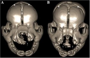
Publication: BMC Vet Res. 2018 Oct 20;14(1):316. PMID: 30342508 | PDF Authors: Pankowski F, Paśko S, Max A, Szal B, Dzierzęcka M, Gruszczyńska J, Szaro P, Gołębiowski M, Bartyzel BJ. Institution: Department of Morphological Sciences, Faculty of Veterinary Medicine, Warsaw University of Life Sciences - SGGW, Warsaw, Poland. Abstract: BACKGROUND: Cleft palate is a birth defect characterized by a lack of fusion between structures forming the palate. Causes include a multitude of factors, both genetic and environmental. Computed tomography (CT) is widely used to evaluate morphological features and diagnose head disorders in adult dogs. However, there is less data about its use in neonatal dogs. The purpose of this study was to perform CT evaluation of palatal defects in one-day-old puppies and to present a novel approach of 3D modeling in terms of cleft palate assessment. RESULTS: Macroscopic and CT examinations were performed in 23 stillborn or euthanized purebred newborn puppies. On the basis of CT data, a 3D model was prepared and the cleft surface area was then calculated. A multi-stage approach, which utilised software such as 3D Slicer and Blender, was applied. Palatal defects were found in ten dogs, of which five had cleft palate, three had bilateral cleft lip and palate, one had a unilateral cleft lip and palate and one had a unilateral cleft lip. The surface area of the clefts ranged from 31 to 213 mm2, which made up respectfully 11 to 63% of the total surface area of the palate. No abnormalities were found in thirteen dogs and they made up the control group. CONCLUSIONS: Computed tomography and 3D modeling were very effective in evaluation of palatal disorders in newborn dogs. 3D models adapted to the natural curvature of the palate were created and more precise data was obtained. Morphological characteristics, CT findings and advanced image analysis of cleft palate in neonates obtained from these models increase the knowledge about this malformation in dogs. |
A Wearable Mixed-reality Holographic Computer for Guiding External Ventricular Drain Insertion at the Bedside
|
Publication: J Neurosurg. 2018 Oct 1:1-8. PMID: 30485188 | PDF Authors: Li Y, Chen X, Wang N, Zhang W, Li D, Zhang L, Qu X, Cheng W, Xu Y, Chen W, Yang Q. Institution: Department of Neurosurgery, Xuanwu Hospital, Capital Medical University, Beijing, China. Abstract: OBJECTIVE: The goal of this study was to explore the feasibility and accuracy of using a wearable mixed-reality holographic computer to guide external ventricular drain (EVD) insertion and thus improve on the accuracy of the classic freehand insertion method for EVD insertion. The authors also sought to provide a clinically applicable workflow demonstration. METHODS: Pre- and postoperative CT scanning were performed routinely by the authors for every patient who needed EVD insertion. Hologram-guided EVD placement was prospectively applied in 15 patients between August and November 2017. During surgical planning, model reconstruction and trajectory calculation for each patient were completed using preoperative CT. By wearing a Microsoft HoloLens, the neurosurgeon was able to visualize the preoperative CT-generated holograms of the surgical plan and perform EVD placement by keeping the catheter aligned with the holographic trajectory. Fifteen patients who had undergone classic freehand EVD insertion were retrospectively included as controls. The feasibility and accuracy of the hologram-guided technique were evaluated by comparing the time required, number of passes, and target deviation for hologram-guided EVD placement with those for classic freehand EVD insertion. "Preoperative DICOM data were imported into 3D Slicer software (version 4.7.0, nightly build, Surgical Planning Laboratory, Harvard Medical School), and segmentation of the lateral ventricles in both groups was performed to calculate the ventricular size." RESULTS: Surgical planning and hologram visualization were performed in all 15 cases in which EVD insertion involved holographic guidance. No adverse events related to the hologram-guided procedures were observed. The mean ± SD additional time before the surgical part of the procedure began was 40.20 ± 10.74 minutes. The average number of passes was 1.07 ± 0.258 in the holographic guidance group, compared with 2.33 ± 0.98 in the control group (p < 0.01). The mean target deviation was 4.34 ± 1.63 mm in the holographic guidance group and 11.26 ± 4.83 mm in the control group (p < 0.01). CONCLUSIONS: This study demonstrates the use of a head-mounted mixed-reality holographic computer to successfully perform hologram-assisted bedside EVD insertion. A full set of clinically applicable workflow images is presented to show how medical imaging data can be used by the neurosurgeon to visualize patient-specific holograms that can intuitively guide hands-on operation. The authors also provide preliminary confirmation of the feasibility and accuracy of this hologram-guided EVD insertion technique. |
 Hologram-guided operation procedures. A: Preoperative CT image of a patient. B: Electrocorticography gel electrodes attached to the head of the patient as registration markers. C and D: Before (C) and after (D) manual rigid coregistration between the holograms and the patient’s head. Arrow denotes hologram of the insertion trajectory. E: The neurosurgeon wore the headset during the whole procedure. F: A burr hole was performed guided by the holographic orientation of the trajectory (arrow). G: The stylet-loaded catheter (upper arrow) insertion was intuitively guided by keeping it aligned with the holographic trajectory (lower arrow). H: Postoperative CT scan verified the accuracy of EVD placement. Arrow denotes the tip of the catheter. Figure is available in color online only. |
The Dark Shades of the Antikythera Mechanism
|
Publication: J Radioanal Nucl Chem. 2018 Oct; | PDF Authors: Aristeidis Voulgaris, Christophoros Mouratidis, Andreas Vossinakis. Institution: Thessaloniki Astronomy Club, Thessaloniki, Greece. Abstract: In this work we analyze the dark shades which are evident on the AMRP X-ray positive Computed Tomographies and Radiographies of the Antikythera Mechanism ancient prototype. During 2000 years under the sea, the Mechanism bronze parts were totally corroded. The decreased X-ray absorption of the corroded bronze, allowed the CTs capture even in the thick large side of Fragment A. During the photometric analysis of the CTs, apart from corroded bronze, at least two other different (corroded) metal materials were detected, which were used by the ancient manufacturer during the construction of the Antikythera Mechanism. From our analysis and correlating with the mechanical evidence resulting by the use of our functional reconstruction models of the Antikythera Mechanism, we conclude that the existing design of the Antikythera Mechanism is probably the first of such a sophisticated design of the ancient manufacturer. |
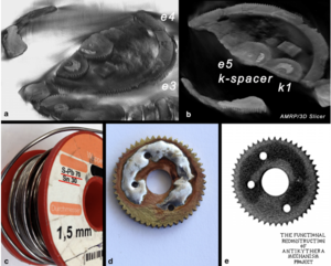 a. 3D reconstruction/visualization of gears e3, e4, e5 and k1, based on the relative AMRP negative tomographies of Fragment A, with the use of 3D Slicer software, processed by the authors. b. A different angle close up view of the 3D reconstruction in the area of k1 gear and k-spacer. Note that just above the spacers there are the gears k2 and e6. c. A typical electronics soldering wire with Pb 70% and Sn 30%. d. Direct stabilization of Pb–Sn solder on a gear by heating. The color of the flux-rosin is also evident. e. The corresponding X-ray radiography @120 keV of the gear on Fig. 6d |
Lung Tumor Segmentation Methods: Impact on the Uncertainty of Radiomics Features for Non-small Cell Lung Cancer
|
Publication: PLoS One. 2018 Oct 4;13(10):e0205003. PMID: 30286184 | PDF Authors: Owens CA, Peterson CB, Tang C, Koay EJ, Yu W, Mackin DS, Li J, Salehpour MR, Fuentes DT, Court LE, Yang J. Institution: Department of Radiation Physics, The University of Texas MD Anderson Cancer Center, Houston, Texas, USA. Abstract: PURPOSE: To evaluate the uncertainty of radiomics features from contrast-enhanced breath-hold helical CT scans of non-small cell lung cancer for both manual and semi-automatic segmentation due to intra-observer, inter-observer, and inter-software reliability. METHODS: Three radiation oncologists manually delineated lung tumors twice from 10 CT scans using two software tools (3D-Slicer and MIM Maestro). Additionally, three observers without formal clinical training were instructed to use two semi-automatic segmentation tools, Lesion Sizing Toolkit (LSTK) and GrowCut, to delineate the same tumor volumes. The accuracy of the semi-automatic contours was assessed by comparison with physician manual contours using Dice similarity coefficients and Hausdorff distances. Eighty-three radiomics features were calculated for each delineated tumor contour. Informative features were identified based on their dynamic range and correlation to other features. Feature reliability was then evaluated using intra-class correlation coefficients (ICC). Feature range was used to evaluate the uncertainty of the segmentation methods. RESULTS: From the initial set of 83 features, 40 radiomics features were found to be informative, and these 40 features were used in the subsequent analyses. For both intra-observer and inter-observer reliability, LSTK had higher reliability than GrowCut and the two manual segmentation tools. All observers achieved consistently high ICC values when using LSTK, but the ICC value varied greatly for each observer when using GrowCut and the manual segmentation tools. For inter-software reliability, features were not reproducible across the software tools for either manual or semi-automatic segmentation methods. Additionally, no feature category was found to be more reproducible than another feature category. Feature ranges of LSTK contours were smaller than those of manual contours for all features. CONCLUSION: Radiomics features extracted from LSTK contours were highly reliable across and among observers. With semi-automatic segmentation tools, observers without formal clinical training were comparable to physicians in evaluating tumor segmentation. "Three radiation oncologists manually delineated lung tumors twice from 10 CT scans using two software tools, 3D Slicer and MIM Maestro." |
 User inputs for initializing semi-automatic segmentation tools (A) LSTK requires the user to select a seed within the tumor (red) to initiate the segmentation algorithm. Defining the maximum tumor radius generates a 3D bounding box (green) centered about the seed, within which the segmentation result will be confined. (B) GrowCut requires the user to label foreground (blue) and background (yellow) pixels to initiate the segmentation algorithm. Once labels were established, the GrowCut algorithm was followed by manual editing of the GrowCut-generated contours. Note that only the transverse view is shown here. Observers also labeled foreground and background pixels in the coronal and sagittal planes for each tumor case. |
Accuracy Assessment of 3D-Printed Tooth Replicas
|
Publication: Curr Med Sci. 2018 Oct;38(5):880-7. PMID: 30341524 Authors: Song P, Duan FL, Cai Q, Wu JL, Chen XB, Wang Y, Huang CG, Li JQ, He ZQ, Huang QC, Liu M, Zhang YG, Luo M. Institution: Department of Neurosurgery, the First Hospital of Wuhan, Wuhan, China. Abstract: The efficacy and applied value of endoscopic hematoma evacuation vs. external ventricular drainage (EVD) in the treatment of severe ventricular hemorrhage (IVH) were explored and compared. From Jan. 2015 to Dec. 2016, the clinical data of 42 cases of IVH were retrospectively analyzed, including 18 patients undergoing endoscopic hematoma evacuation (group A), and 24 patients receiving EVD (group B). The hematoma clearance rate was calculated by 3D Slicer software, and complications and outcomes were compared between the two groups. There were no significant differences in age, sex and Graeb score between groups A and B (P>0.05). The hematoma clearance rate was 70.81%±27.64% in group A and 48.72%±36.58% in group B with a statistically significant difference (P<0.05). The operative time in groups A and B was 72.45±25.26 min and 28.54±15.27 min, respectively (P<0.05). The Glasgow Coma Scale (GCS) score increased from 9.28±2.72 at baseline to 11.83±2.91 at 1 week postoperatively in group A, and from 8.25±2.62 at baseline to 10.79±4.12 at 1 week postoperatively in group B (P<0.05). The length of hospital stay was 12.67±5.97 days in group A and 17.33±8.91 days in group B with a statistically significant difference (P<0.05). The GOS scores at 6 months after surgery were 3.83±1.12 in group A, and 2.75±1.23 in group B (P<0.05). These results suggested that endoscopic hematoma evacuation has an advantage of a higher hematoma clearance rate, fewer complications and better outcomes in the treatment of severe IVH, indicating it is a safe, effective and promising approach for severe IVH. |
Non-invasive Magnetic Resonance Imaging of Oils in Botryococcus Braunii Green Algae: Chemical Shift Selective and Diffusion-weighted Imaging
|
Publication: PLoS One. 2018 Oct 4;13(10):e0205003. PMID: 30161202 | PDF Authors: Schadewijk RV, Berg TEVD, Gupta KBSS, Ronen I, de Groot HJM, Alia A. Institution: Leiden Institute of Chemistry, Leiden University, Leiden, The Netherlands. Abstract: Botryococcus braunii is an oleaginous green algae with the distinctive property of accumulating high quantities of hydrocarbons per dry weight in its colonies. Large variation in colony structure exists, yet its implications and influence of oil distribution and diffusion dynamics are not known and could not be answered due to lack of suitable in vivo methods. This publication seeks to further the understanding on oil dynamics, by investigating naturally relevant large (700-1500μm) and extra-large (1500-2500μm) sized colonies of Botryococcus braunii (race B, strain Showa) in vivo, using a comprehensive approach of chemical shift selective imaging, chemical shift imaging and spin echo diffusion measurements at high magnetic field (17.6T). Hydrocarbon distribution in large colonies was found to be localised in two concentric oil layers with different thickness and concentration. Extra-large colonies were highly unstructured and oil was spread throughout colonies, but with large local variations. Interestingly, fluid channels were observed in extra-large colonies. Diffusion-weighted MRI revealed a strong correlation between colony heterogeneity, oil distribution, and diffusion dynamics in different parts of Botryococcus colonies. Differences between large and extra-large colonies were characterised by using T2 weighted MRI along with relaxation measurements. Our result, therefore, provides first non-invasive MRI means to obtain spatial information on oil distribution and diffusion dynamics in Botryococcus braunii colonies. "3D-MSME data were exported to DICOM using Paravision 5.1, consequently reconstructed in 3D Slicer v.4.8..." |
 T1 and T2 relaxometry and mapping of B. braunii colonies. Relaxation measurement was performed using RAREVTR sequence (TR-array, 5500–200 ms; TE, 27–4.5 ms; number of averages, 16; matrix size, 128 x 128; FOV, 0.5 x 0.5mm; resolution was 39 x 39 x 250 μm3). (A) A representative image showing regions of interest (ROI) placed on two representative colonies (one large and one extra-large size colony) for calculating T1 and T2 relaxation times. Scale bar: 500 μm. (B) T1 Map derived from RAREVTR sequence, showing the region of high (white arrow) and low T1 (white arrowhead). Colour scale was generated with Paravision ‘colour 256’ scheme which ranges from 0 to 2500ms. (C) T2 Map Derived from RAREVTR sequence showing a sharp edge of low T2 surrounding all colonies (black arrow). Colour scale ranges from 0 to 40ms. |
Altered White Matter Connectivity in Patients with Schizophrenia: An Investigation using Public Neuroimaging Data from SchizConnect
|
Publication: PLoS One. 2018 Oct 9;13(10):e0205369. PMID: 30300425 | PDF Authors: Joo SW, Yoon W, Shon SH, Kim H, Cha S, Park KJ, Lee J. Institution: Department of Psychiatry, Asan Medical Center, University of Ulsan College of Medicine, Seoul, Korea. Abstract: Several studies have produced extensive evidence on white matter abnormalities in schizophrenia (SZ). However, optimum consistency and reproducibility have not been achieved, and reported low white matter tract integrity in patients with SZ varies between studies. A whole-brain imaging study with a large sample size is needed. This study aimed to investigate white matter integrity in the corpus callosum and connections between regions of interests (ROIs) in the same hemisphere in 122 patients with SZ and 129 healthy controls with public neuroimaging data from SchizConnect. For each diffusion-weighted image (DWI), two-tensor full-brain tractography was performed; DWIs were parcellated by processing and registering T1 images with FreeSurfer and Advanced Normalization Tools. White matter query language was used to extract white matter fiber tracts. We evaluated group differences in means of diffusion measures between the patients and controls, and correlations of diffusion measures with the severity of clinical symptoms and cognitive impairment in the patients using the Positive and Negative Syndrome Scale (PANSS), a letter-number sequencing (LNS) test, vocabulary test, letter fluency test, category fluency test, and trail-making test, part A. To correct for multiple comparisons, a false discovery rate of q < 0.05 was applied. In patients with SZ, we observed significant radial diffusivity (RD) and trace (TR) increases in left thalamo-occipital tracts and the right uncinate fascicle, and a significant RD increase in the right middle longitudinal fascicle (MDLF) and the right superior longitudinal fascicle ii. Correlations were present between TR of left thalamo-occipital tracts, and the letter fluency test and the LNS test, and RD in the right MDLF and PANSS positive subscale score. However, these correlations were not significant after correction for multiple comparisons. These results indicated widespread white matter fiber tract abnormalities in patients with SZ, contributing to SZ pathophysiology. "We used UKF tractography implemented in the 3D Slicer to perform whole-brain tractography" |
SlicerSALT: Shape AnaLysis Toolbox
|
Publication: Shape Med Imaging (2018). 2018 Sep;11167:65-72 PMID: 31032495 | PDF Authors: Vicory J, Pascal L, Hernandez P, Fishbaugh J, Prieto J, Mostapha M, Huang C, Shah H, Hong J, Liu Z, Michoud L, Fillion-Robin JC, Gerig G, Zhu H, Pizer SM, Styner M, Paniagua B. Institution: Kitware, Inc. Abstract: SlicerSALT is an open-source platform for disseminating state-of-the-art methods for performing statistical shape analysis. These methods are developed as 3D Slicer extensions to take advantage of its powerful underlying libraries. SlicerSALT itself is a heavily customized 3D Slicer package that is designed to be easy to use for shape analysis researchers. The packaged methods include powerful techniques for creating and visualizing shape representations as well as performing various types of analysis. Funding:
|
Association of Anemia and Hemoglobin Decrease during Acute Stroke Treatment with Infarct Growth and Clinical Outcome
|
Publication: PLoS One. 2018 Sep 26;13(9):e0203535. PMID: 30256814 | PDF Authors: Bellwald S, Balasubramaniam R, Nagler M, Burri MS, Fischer SDA, Hakim A, Dobrocky T, Yu Y, Scalzo F, Heldner MR, Wiest R, Mono ML, Sarikya H, El-Koussy M, Mordasini P, Fischer U, Schroth G, Gralla J, Mattle HP, Arnold M, Liebeskind D, Jung S. Institution: Department of Neurology, Inselspital, Bern University Hospital, University of Bern, Bern, Switzerland. Abstract: BACKGROUND AND PURPOSE: Anemia is associated with worse outcome in stroke, but the impact of anemia with intravenous thrombolysis or endovascular therapy has hardly been delineated. The aim of this study was to analyze the role of anemia on infarct evolution and outcome after acute stroke treatment. METHODS: 1158 patients from Bern and 321 from Los Angeles were included. Baseline data and 3 months outcome assessed with the modified Rankin Scale were recorded prospectively. Baseline DWI lesion volumes were measured in 345 patients and both baseline and final infarct volumes in 180 patients using CT or MRI. Multivariable and linear regression analysis were used to determine predictors of outcome and infarct growth. RESULTS: 712 patients underwent endovascular treatment and 446 intravenous thrombolysis. Lower hemoglobin at baseline, at 24h, and nadir until day 5 predicted poor outcome (OR 1.150-1.279) and higher mortality (OR 1.131-1.237) independently of treatment. Decrease of hemoglobin after hospital arrival, mainly induced by hemodilution, predicted poor outcome and had a linear association with final infarct volumes and the amount and velocity of infarct growth. Infarcts of patients with newly observed anemia were twice as large as infarcts with normal hemoglobin levels. CONCLUSION: Anemia at hospital admission and any hemoglobin decrease during acute stroke treatment affect outcome negatively, probably by enlarging and accelerating infarct growth. Our results indicate that hemodilution has an adverse effect on penumbral evolution. Whether hemoglobin decrease in acute stroke could be avoided and whether this would improve outcome would need to be studied prospectively. "Segmentation of the final infarct was performed with 3D Slicer v.4.5.0." |
Outer Wall Segmentation of Abdominal Aortic Aneurysm by Variable Neighborhood Search Through Intensity and Gradient Spaces
|
Publication: J Digit Imaging. 2018 Aug;31(4):490-504. PMID: 29352385 | PDF Authors: Siriapisith T, Kusakunniran W, Haddawy P2. Institution: Department Radiology, Faculty of Medicine Siriraj Hospital, Mahidol University, Bangkok, Thailand. Abstract: Aortic aneurysm segmentation remains a challenge. Manual segmentation is a time-consuming process which is not practical for routine use. To address this limitation, several automated segmentation techniques for aortic aneurysm have been developed, such as edge detection-based methods, partial differential equation methods, and graph partitioning methods. However, automatic segmentation of aortic aneurysm is difficult due to high pixel similarity to adjacent tissue and a lack of color information in the medical image, preventing previous work from being applicable to difficult cases. This paper uses uses a variable neighborhood search that alternates between intensity-based and gradient-based segmentation techniques. By alternating between intensity and gradient spaces, the search can escape from local optima of each space. The experimental results demonstrate that the proposed method outperforms the other existing segmentation methods in the literature, based on measurements of dice similarity coefficient and jaccard similarity coefficient at the pixel level. In addition, it is shown to perform well for cases that are difficult to segment. "Ground truth segmentations were also obtained by manual segmentation with the 3D Slicer version 4.7.0." |
 An example of graph cut with probability density function corresponding to the diagram in Fig. 5. a. The initial contour for the segmentation. b. The label region with bone and air removal. For the graph construction, each pixel in the region is represented by a node in the graph. c. The output label region of segmentation using max-flow/min-cut algorithm. d. The output contour of segmentation corresponds to the outer boundary of c. |
Simple and Robust Referencing System Enables Identification of Dissolved-phase Xenon Spectral Frequencies
|
Publication: Magn Reson Med. 2018 Aug;80(2):431-41. PMID: 29266425 | PDF Authors: Antonacci MA, Zhang L, Burant A, McCallister D, Branca RT. Institution: Department of Physics and Astronomy, University of North Carolina at Chapel Hill, NC, USA. Abstract: To assess the effect of macroscopic susceptibility gradients on the gas-phase referenced dissolved-phase 129 Xe (DPXe) chemical shift (CS) and to establish the robustness of a water-based referencing system for in vivo DPXe spectra. METHODS: Frequency shifts induced by spatially varying magnetic susceptibility are calculated by finite-element analysis for the human head and chest. Their effect on traditional gas-phase referenced DPXe CS is then assessed theoretically and experimentally. A water-based referencing system for the DPXe resonances that uses the local water protons as reference is proposed and demonstrated in vivo in rats. RESULTS: Across the human brain, macroscopic susceptibility gradients can induce an apparent variation in the DPXe CS of up to 2.5 ppm. An additional frequency shift as large as 6.5 ppm can exist between DPXe and gas-phase resonances. By using nearby water protons as reference for the DPXe CS, the effect of macroscopic susceptibility gradients is eliminated and consistent CS values are obtained in vivo, regardless of shimming conditions, region of interest analyzed, animal orientation, or lung inflation. Combining in vitro and in vivo spectroscopic measurements finally enables confident assignment of some of the DPXe peaks observed in vivo. CONCLUSION: To use hyperpolarized xenon as a biological probe in tissues, the DPXe CS in specific organs/tissues must be reliably measured. When the gas-phase is used as reference, variable CS values are obtained for DPXe resonances. Reliable peak assignments in DPXe spectra can be obtained by using local water protons as reference. "Each region was then assembled into a standard tessellation language mesh using 3D Slicer." |
 (a) Representative maps (left column) of the frequency shift induced by magnetic susceptibility gradients at 3 T in the human head along with (right column) corresponding original CT slices used in the model. In CT slices, white represents bones, gray represents soft tissue, and black represents air. (b) Histograms of frequency shift (left) present within the highlighted voxels shown in the 3D human head rendering (right). Note that anatomical features of the model located in front of the planes and voxels of interest are visible in the model rendering. |
Diffusion Weighted And Dynamic Contrast Enhanced MRI as an Imaging Biomarker for Stereotactic Ablative Body Radiotherapy (SABR) of Primary Renal Cell Carcinoma
|
Publication: PLoS One. 2018 Aug 16;13(8):e0202387. PMID: 30114235 | PDF Authors: Reynolds HM, Parameswaran BK, Finnegan ME, Roettger D, Lau E, Kron T, Shaw M, Chander S, Siva S. Institution: Department of Physical Sciences, Peter MacCallum Cancer Centre, Melbourne, Victoria, Australia. Abstract: PURPOSE: To explore the utility of diffusion and perfusion changes in primary renal cell carcinoma (RCC) after stereotactic ablative body radiotherapy (SABR) as an early biomarker of treatment response, using diffusion weighted (DWI) and dynamic contrast enhanced (DCE) MRI. METHODS: Patients enrolled in a prospective pilot clinical trial received SABR for primary RCC, and had DWI and DCE MRI scheduled at baseline, 14 days and 70 days after SABR. Tumours <5cm diameter received a single fraction of 26 Gy and larger tumours received three fractions of 14 Gy. Apparent diffusion coefficient (ADC) maps were computed from DWI data and parametric and pharmacokinetic maps were fitted to the DCE data. Tumour volumes were contoured and statistics extracted. Spearman's rank correlation coefficients were computed between MRI parameter changes versus the percentage tumour volume change from CT at 6, 12 and 24 months and the last follow-up relative to baseline CT. RESULTS: Twelve patients were eligible for DWI analysis, and a subset of ten patients for DCE MRI analysis. DCE MRI from the second follow-up MRI scan showed correlations between the change in percentage voxels with washout contrast enhancement behaviour and the change in tumour volume (ρ = 0.84, p = 0.004 at 12 month CT, ρ = 0.81, p = 0.02 at 24 month CT, and ρ = 0.89, p = 0.001 at last follow-up CT). The change in mean initial rate of enhancement and mean Ktrans at the second follow-up MRI scan were positively correlated with percent tumour volume change at the 12 month CT onwards (ρ = 0.65, p = 0.05 and ρ = 0.66, p = 0.04 at 12 month CT respectively). Changes in ADC kurtosis from histogram analysis at the first follow-up MRI scan also showed positive correlations with the percentage tumour volume change (ρ = 0.66, p = 0.02 at 12 month CT, ρ = 0.69, p = 0.02 at last follow-up CT), but these results are possibly confounded by inflammation. CONCLUSION: DWI and DCE MRI parameters show potential as early response biomarkers after SABR for primary RCC. Further prospective validation using larger patient cohorts is warranted. |
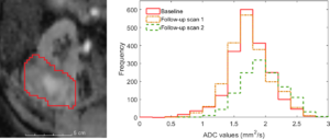 Baseline ADC map with tumour contour in red for patient 3 and associated histogram showing the frequency of ADC values in each MRI scan. ADC maps were read into 3D Slicer software and the tumour volume contoured directly onto ADC map image slices by an experienced radiologist. |
Toward a Real-time System for Temporal Enhanced Ultrasound-guided Prostate Biopsy
|
Publication: Int J Comput Assist Radiol Surg. 2018 Aug;13(8):1201-9. PMID: 29589258 | PDF Authors: Azizi S, Van Woudenberg N, Sojoudi S, Li M, Xu S, Abu Anas EM, Yan P, Tahmasebi A, Kwak JT, Turkbey B, Choyke P, Pinto P, Wood B, Mousavi P, Abolmaesumi P. Institution: The University of British Columbia, Vancouver, BC, Canada. Abstract: PURPOSE: We have previously proposed temporal enhanced ultrasound (TeUS) as a new paradigm for tissue characterization. TeUS is based on analyzing a sequence of ultrasound data with deep learning and has been demonstrated to be successful for detection of cancer in ultrasound-guided prostate biopsy. Our aim is to enable the dissemination of this technology to the community for large-scale clinical validation. METHODS: In this paper, we present a unified software framework demonstrating near-real-time analysis of ultrasound data stream using a deep learning solution. The system integrates ultrasound imaging hardware, visualization and a deep learning back-end to build an accessible, flexible and robust platform. A client-server approach is used in order to run computationally expensive algorithms in parallel. We demonstrate the efficacy of the framework using two applications as case studies. First, we show that prostate cancer detection using near-real-time analysis of RF and B-mode TeUS data and deep learning is feasible. Second, we present real-time segmentation of ultrasound prostate data using an integrated deep learning solution. RESULTS: The system is evaluated for cancer detection accuracy on ultrasound data obtained from a large clinical study with 255 biopsy cores from 157 subjects. It is further assessed with an independent dataset with 21 biopsy targets from six subjects. In the first study, we achieve area under the curve, sensitivity, specificity and accuracy of 0.94, 0.77, 0.94 and 0.92, respectively, for the detection of prostate cancer. In the second study, we achieve an AUC of 0.85. CONCLUSION: Our results suggest that TeUS-guided biopsy can be potentially effective for the detection of prostate cancer. |
 Guidance interface implemented as part of a 3D Slicer module: cancer likelihood map is overlaid on B-mode ultrasound images. Red indicates predicted labels as cancer, and blue indicates predicted benign regions. The boundary of the segmented prostate is shown with white, and the green circle is centered around the target location which is shown in green dot. |
Innovations in Preoperative Planning: Insights into Another Dimension using 3D Printing for Cardiac Disease
|
Publication: J Cardiothorac Vasc Anesth. 2018 Aug;32(4):1937-45. PMID: 29277300 Authors: Farooqi KM, Mahmood F. Institution: Division of Pediatric Cardiology, New York Presbyterian-Columbia University Medical Center, New York, NY, USA. Abstract: Two-dimensional visualization of complex congenital heart disease has limitations in that there is variation in the interpretation by different individuals. Three-dimensional printing technology has been in use for decades but is currently becoming more commonly used in the medical field. Congenital heart disease serves as an ideal pathology to employ this technology because of the variation of anatomy between patients. In this review, the authors aim to discuss basics of applicability of three-dimensional printing, the process involved in creating a model, as well as challenges with establishing utility and quality. |
3D-constructive Interference into Steady State (3D-CISS) Labyrinth Signal Alteration in Patients with Vestibular Schwannoma
|
Publication: Auris Nasus Larynx. 2018 Aug;45(4):702-10. PMID: 28947096 Authors: Wagner F, Herrmann E, Wiest R, Raabe A, Bernasconi C, Caversaccio M, Vibert D. Institutions: Department of Diagnostic and Interventional Neuroradiology, University Hospital, University of Bern, Bern, Switzerland. Abstract: Objective: To evaluate signal intensity of the inner ear using 3D-CISS imaging and correlated signal characteristics in patients with vestibular schwannoma to neuro-otological symptoms. Methods: Sixty patients with unilateral vestibular schwannoma were retrospectively reviewed. All patients had had initial and follow-up magnetic resonance imaging (MRI). Individual treatment strategies consisted of "wait-and-watch", surgical tumour resection, stereotactic radiosurgery or both surgery and stereotactic radiosurgery. For all patients a complete baseline and treatment course neuro-otological examination was re-studied. For the semi-automatic volumetric tumour measurement we used 3D Slicer 4.4.0. Results: On initial MRI, 3D-CISS sequence signal loss of the membranous labyrinth was present in 20 patients (33.3%); signal loss of cochlea in 20 (33.3%) and coincident signal loss of sacculus/utriculus in 17 (85%) of them. Sequential analysis of follow-up MRI series demonstrated slightly increased labyrinthine signal degradation, independently of the chosen therapy. Correlation of initial MRI results with initial neuro-otological symptoms showed significance only for cochlear obstruction versus vertigo (p=0.0397) and sacculus/utriculus obstruction versus vertigo (p=0.0336). No other statistically significant relationships were noted. Conclusion: 3D-constructive interference into steady state (3D-CISS) is appropriate for observing inner ear signal loss in patients with vestibular schwannoma. However, except for vertigo, no significant correlation was noted between initial neuro-otological symptomatology and signal loss of the inner ear. |
Computed Tomography Trachea Volumetry in Patients With Scleroderma: Association With Clinical and Functional Findings
|
Publication: PLoS One. 2018 Aug 1;13(8):e0200754. PMID: 30067820 | PDF Authors: Silva BRA, Rodrigues RS, Rufino R, Costa CH, Vilela VS, Levy RA, Guimarães ARM, Carvalho ARS, Lopes AJ. Institution: Postgraduate Programme in Medical Sciences, School of Medical Sciences, State University of Rio de Janeiro, Rio de Janeiro, Brazil. Abstract: BACKGROUND: In scleroderma, excessive collagen production can alter tracheal geometry, and computed tomography (CT) volumetry of this structure may aid in detecting possible abnormalities. The objectives of this study were to quantify the morphological abnormalities in the tracheas of patients with scleroderma and to correlate these findings with data on clinical and pulmonary function. METHODS: This was a cross-sectional study in which 28 adults with scleroderma and 27 controls matched by age, gender and body mass index underwent chest CT with posterior segmentation and skeletonization of the images. In addition, all participants underwent pulmonary function tests and clinical evaluation, including the modified Rodnan skin score (mRSS). RESULTS: Most patients (71.4%) had interstitial lung disease on CT. Compared to controls, patients with scleroderma showed higher values in the parameters measured by CT trachea volumetry, including area, eccentricity, major diameter, minor diameter, and tortuosity. The tracheal area and equivalent diameter were negatively correlated with the ratio between forced expiratory flow and forced inspiratory flow at 50% of forced vital capacity (FEF50%/FIF50%) (r = -0.44, p = 0.03 and r = -0.46, p = 0.02, respectively). The tracheal tortuosity was negatively correlated with peak expiratory flow (r = -0.51, p = 0.008). The mRSS showed a positive correlation with eccentricity (r = 0.62, p < 0.001) and tracheal tortuosity (r = 0.51, p = 0.007), while the presence of anti-topoisomerase I antibody (ATA) showed a positive correlation with tracheal tortuosity (r = 0.45, p = 0.03). CONCLUSIONS: In a sample composed predominantly of scleroderma patients with associated interstitial lung disease, there were abnormalities in tracheal geometry, including greater eccentricity, diameter and tortuosity. In these patients, abnormalities in the geometry of the trachea were associated with functional markers of obstruction. In addition, tracheal tortuosity was correlated with cutaneous involvement and the presence of ATA. |
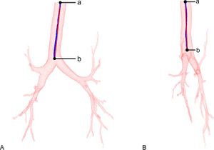 Measurement of the tortuosity of the trachea. Images in the coronal (A) and sagittal (B) planes are shown. In this scheme, L is the length of the trachea (the total length of 'ab', shown in blue) and vd is the vectorial distance between the points at the extremities (the length of the shortest possible path between 'a’ and ‘b', shown in red).The airways were segmented using 3D Slicer v.4.4.0 with the aid of its AirwaySegmentation extension. |
Comparison of Modified Two-point Dixon and Chemical Shift Encoded MRI Water-fat Separation Methods for Fetal Fat Quantification
|
Publication: J Magn Reson Imaging. 2018 Jul;48(1):274-82. PMID: 29319918 Authors: Giza SA, Miller MR, Parthasarathy P, de Vrijer B, McKenzie CA. Institution: Department of Medical Biophysics, Western University, London, Ontario, Canada. Abstract: BACKGROUND: Fetal fat is indicative of the energy balance within the fetus, which may be disrupted in pregnancy complications such as fetal growth restriction, macrosomia, and gestational diabetes. Water-fat separated MRI is a technique sensitive to tissue lipid content, measured as fat fraction (FF), and can be used to accurately measure fat volumes. Modified two-point Dixon and chemical shift encoded MRI (CSE-MRI) are water-fat separated MRI techniques that could be applied to imaging of fetal fat. Modified two-point Dixon has biases present that are corrected in CSE-MRI which may contribute to differences in the fat measurements. PURPOSE: To compare the measurement of fetal fat volume and FF by modified two-point Dixon and CSE-MRI. STUDY TYPE: Cross-sectional study for comparison of two MRI pulse sequences. POPULATION: Twenty-one pregnant women with singleton pregnancies. FIELD STRENGTH/SEQUENCE: 1.5T, modified two-point Dixon and CSE-MRI. ASSESSMENT: Manual segmentation of total fetal fat volume and mean FF from modified 2-point Dixon and CSE-MRI FF images. STATISTICAL TESTS: Reliability was assessed by calculating the intraclass correlation coefficient (ICC). Agreement was assessed using a one-sample t-test on the fat measurements difference values (modified two-point Dixon - CSE-MRI). The difference scores were tested against a value of 0, which would indicate that the measurements were identical. RESULTS: The fat volume and FF measured by modified two-point Dixon and CSE-MRI had excellent reliability, demonstrated by ICCs of 0.93 (P < 0.001) and 0.90 (P < 0.001), respectively. They were not in agreement, with CSE-MRI giving mean fat volumes 180 mL greater and mean FF 3.0% smaller than modified two-point Dixon. DATA CONCLUSION: The reliability between modified two-point Dixon and CSE-MRI indicates that either technique can be used to compare fetal fat measurements in different participants, but they are not in agreement possibly due to uncorrected biases in modified two-point Dixon. "Total fetal fat from the entire fetal volume was manually segmented from all the PDFF/FSF images using 3D Slicer v.4.7.0." |
Genetic Mapping of Molar Size Relations Identifies Inhibitory Locus for Third Molars in Mice
|
Publication: Heredity (Edinb). 2018 Jul;121(1):1-11. PMID: 29302051 | PDF Authors: Navarro N, Murat Maga A. Institution: EPHE, PSL Research University Paris, F-21000, Dijon, France. Abstract: Molar size in Mammals shows considerable disparity and exhibits variation similar to that predicted by the Inhibitory Cascade model. The importance of such developmental systems in favoring evolutionary trajectories is also underlined by the fact that this model can predict macroevolutionary patterns. using backcross mice, we mapped QTL for molar sizes controlling for their sequential development. Genetic controls for upper and lower molars appear somewhat similar, and regions containing genes implied in dental defects drive this variation. We mapped three relationship QTLs (rQTL) modifying the control of the mesial molars on the focal third molar. These regions overlap Shh, Sostdc1, and Fst genes, which have pervasive roles in development and should be buffered against new variation. It has theoretically been shown that rQTL produces new variation channeled in the direction of adaptive changes. Our results provide evidence that evolutionary/disease patterns of tooth size variation could result from such a non-random generating process. "A random set of 79 individuals was segmented using the 3D Slicer with a specific threshold."
|
Characterization of Adrenal Lesions on Unenhanced MRI Using Texture Analysis: A Machine-Learning Approach
|
Publication: J Magn Reson Imaging. 2018 Jul;48(1):198-204. PMID: 29341325 Authors: Romeo V, Maurea S, Cuocolo R, Petretta M, Mainenti PP, Verde F, Coppola M, Dell'Aversana S, Brunetti A. Institution: Department of Advanced Biomedical Sciences, University of Naples "Federico II,", Naples, Italy. Abstract: BACKGROUND: Adrenal adenomas (AA) are the most common benign adrenal lesions, often characterized based on intralesional fat content as either lipid-rich (LRA) or lipid-poor (LPA). The differentiation of AA, particularly LPA, from nonadenoma adrenal lesions (NAL) may be challenging. Texture analysis (TA) can extract quantitative parameters from MR images. Machine learning is a technique for recognizing patterns that can be applied to medical images by identifying the best combination of TA features to create a predictive model for the diagnosis of interest. PURPOSE/HYPOTHESIS: To assess the diagnostic efficacy of TA-derived parameters extracted from MR images in characterizing LRA, LPA, and NAL using a machine-learning approach. STUDY TYPE: Retrospective, observational study. POPULATION/SUBJECTS/PHANTOM/SPECIMEN/ANIMAL MODEL: Sixty MR examinations, including 20 LRA, 20 LPA, and 20 NAL. FIELD STRENGTH/SEQUENCE: Unenhanced T1 -weighted in-phase (IP) and out-of-phase (OP) as well as T2 -weighted (T2 -w) MR images acquired at 3T. ASSESSMENT: Adrenal lesions were manually segmented, placing a spherical volume of interest on IP, OP, and T2 -w images. Different selection methods were trained and tested using the J48 machine-learning classifiers. STATISTICAL TESTS: The feature selection method that obtained the highest diagnostic performance using the J48 classifier was identified; the diagnostic performance was also compared with that of a senior radiologist by means of McNemar's test. RESULTS: A total of 138 TA-derived features were extracted; among these, four features were selected, extracted from the IP (Short_Run_High_Gray_Level_Emphasis), OP (Mean_Intensity and Maximum_3D_Diameter), and T2 -w (Standard_Deviation) images; the J48 classifier obtained a diagnostic accuracy of 80%. The expert radiologist obtained a diagnostic accuracy of 73%. McNemar's test did not show significant differences in terms of diagnostic performance between the J48 classifier and the expert radiologist. DATA CONCLUSION: Machine learning conducted on MR TA-derived features is a potential tool to characterize adrenal lesions. "Images and VOIs were successively imported on 3D Slicer (HeterogeneityCAD module) to extract a total of 138 first-order, GLCM, and RLM texture parameters, 46 for each MR sequence. |
Role of CT and MRI in the Design and Development of Orthopaedic Model using Additive Manufacturing
|
Publication: J Clin Orthop Trauma. 2018 Jul-Sep;9(3):213-7. PMID: 30202151 | PDF Authors: Haleem A, Javaid M. Institution: Department of Mechanical Engineering, Jamia Millia Islamia, New Delhi, India. Abstract: OBJECTIVE: To study the role of Computed tomography (CT) and Magnetic resonance imaging (MRI) for design and development of orthopaedic model using additive manufacturing (AM) technologies. METHODS: A significant number of research papers in this area are studied to provide the direction of development along with the future scope. RESULTS: Briefly discussed various steps used to create a 3D model by Additive Manufacturing using CT and MRI scan. These scanning technologies are used to produce medical as well as orthopaedic implants by using AM technologies. The images so produced are exported in different software like OsiriX Imaging Software, 3D slicer, Mimics, Magics, 3D doctor and InVesalius to produce a 3D digital model. Various criteria's achieved by CT and MRI scan for design and development of orthopaedic implant using additive manufacturing are also discussed briefly. AM model created by this process show exact shape, size, dimensions, textures, colour and features. CONCLUSION: AM technologies help to convert the digital model into a 3D physical object, thereby improving the understanding of patient anatomy for treatment as well as for educational purpose. These scanning technologies have various applications to enhance the AM in the field of orthopaedic. In orthopaedic every patient model is a customised unit, sourced from the individual patient. 3D CAD data captured by these scanning technologies are directly exported in standard triangulate language (STL) format for printing by AM technologies. Crossestion of the physical model fabricated by this process shows a patient's anatomy if the model prepared by using the bone-like material. "The images so produced are exported in different software like OsiriX Imaging Software, 3D Slicer, Mimics, Magics, 3D doctor and InVesalius to produce a 3D digital model. |
Comparing Damage on Retrieved Total Elbow Replacement Bushings with Lab Worn Specimens Subjected to Varied Loading Conditions
|
Publication: J Orthop Res. 2018 Jul;36(7):1998-2006. PMID: 29315772 | PDF Authors: Willing R. Institution: Department of Mechanical Engineering, Thomas J. Watson School of Engineering and Applied Science, State University of New York at Binghamton, Binghamton, NY, USA. Abstract: Complication rates following total elbow replacement (TER) with conventional implants are relatively high due to mechanical failure involving the UHMWPE bushings. Unfortunately, there are no standardized pre-clinical durability testing protocols for assessing the durability of TER components. This study examines the damage observed on retrieved humeral bushings, and then uses in vitro durability testing with two different loading protocols to compare resulting damage. Damage on 25 pairs of retrieved humeral bushings was characterized using micro-computed tomographic imaging techniques. The damage was compared with that of in vitro test specimens which were subjected to 200K cycles of either high joint reaction force (high JRF) or high varus moment (high VM) loading. Material removal (mass loss) from bushing components was measured using gravimetric techniques. Thinning was less for retrieved bushings which were still assembled in their humeral component, versus bushings which were loose (0.3 ± 0.3 mm versus 0.6 ± 0.3 mm, p = 0.02). Comparing in vitro test specimens, thinning due to high VM loading was 0.9 ± 0.3 mm, versus 0.2 ± 0.0 mm for high JRF loading (p = 0.08); however, the actual material removal rates from the humeral bushings were not different between the two protocols (48 ± 5 mm3 /Mc versus 43 ± 2 mm3 /Mc, p = 1). Neither loading protocol could produce damage patterns fully representative of the spectrum of damage patterns observed on clinical retrievals. Pre-clinical testing should employ multiple loading protocols to characterize implant performance under a broader spectrum of usage. This article is protected by copyright. All rights reserved. "DICOM files were imported into 3D Slicer for three-dimensional (3D) model reconstruction." |
Validation of a Windowing Protocol for Accurate In Vivo Tooth Segmentation Using i-Cat Cone Beam Computed Tomography
|
Publication: Adv Clin Exp Med. 2018 Jul;27(7):1001-8. PMID: 29999253 | PDF Authors: Rastegar B, Thumilaire B, Odri GA, Siciliano S, Zapała J, Mahy P, Olszewski R. Institution: Service de Parodontologie, Université libre de Bruxelles, Brussels, Belgium. Abstract: BACKGROUND: Validation of three-dimensional (3D) reconstructions of full dental arches with crowns and roots based on cone beam computed tomography (CBCT) imaging represents a key issue in 3D digital dentistry. OBJECTIVES: The aim of the study was to search for the most accurate in vivo windowing-based manual tooth segmentation using CBCT. The null hypothesis was that all applied windowing protocols were equivalent in terms of in vivo tooth volume measurement using CBCT. MATERIAL AND METHODS: This retrospective study was based on preoperative CBCT images from patients who underwent further tooth extractions for reasons independent of this study. Written informed consent was obtained from all the participants, and the study was approved by the Ethics Committee of Cliniques Universitaires Saint Luc (Brussels, Belgium). The radiological protocol was I-CAT CBCT, 0.3 mm slice thickness, 8 cm × 16 cm field of view, 120 kVp, and 18 mAs. A total of 36 teeth were extracted from 14 patients between the ages of 18 and 68 years. Using 3D Slicer software, segmentations were performed twice by 2 independent observers, with a 1-month time period between the 2 segmentations to study intra and inter-observer repeatability and reproducibility. Four windowing protocols (level/window) were applied: 1. 1131/1858, 2. 2224/4095, 3. 1131/4095, and 4. AUTO, an automatic protocol provided by default by the software. A total of 576 segmentations were performed. Tooth volumes were automatically calculated using the software. To compare the volumes obtained from CBCT segmentations with a gold-standard method, we laser-scanned the extracted teeth. RESULTS: Excellent intra and inter-observer intraclass correlations were found for all of the protocols used. The best windowing protocol was 1131/1858 for both observers. Tooth volumes were obtained by manual segmentation of the CBCT images and using windowing protocol 1131/1858. No significantly different tooth volumes were found by laser scanning. CONCLUSIONS: Our null hypothesis was rejected. Only windowing protocol 1131/1858 allowed for significantly closer 3D in vivo segmentation of a tooth compared to I-CAT CBCT, with excellent intra-observer repeatability and inter-observer reproducibility. |
 Manual segmentation of tooth number 11 with 3D Slicer software A – segmentation on axial slice B – segmentation on sagittal slice C – three-dimensional reconstruction and automatic volume measurement. |
Automatic Segmentation of Stereoelectroencephalography (SEEG) Electrodes Post-Implantation Considering Bending
|
Publication: Int J Comput Assist Radiol Surg. 2018 Jun;13(6):935-46. PMID: 29736800 | PDF Authors: Granados A, Vakharia V, Rodionov R, Schweiger M, Vos SB, O'Keeffe AG, Li K, Wu C, Miserocchi A, McEvoy AW, Clarkson MJ, Duncan JS, Sparks R, Ourselin S. Institution: Wellcome/EPSRC Centre for Interventional and Surgical Sciences, UCL, London, UK. Abstract: PURPOSE: The accurate and automatic localisation of SEEG electrodes is crucial for determining the location of epileptic seizure onset. We propose an algorithm for the automatic segmentation of electrode bolts and contacts that accounts for electrode bending in relation to regional brain anatomy. METHODS: Co-registered post-implantation CT, pre-implantation MRI, and brain parcellation images are used to create regions of interest to automatically segment bolts and contacts. Contact search strategy is based on the direction of the bolt with distance and angle constraints, in addition to post-processing steps that assign remaining contacts and predict contact position. We measured the accuracy of contact position, bolt angle, and anatomical region at the tip of the electrode in 23 post-SEEG cases comprising two different surgical approaches when placing a guiding stylet close to and far from target point. Local and global bending are computed when modelling electrodes as elastic rods. RESULTS: Our approach executed on average in 36.17 s with a sensitivity of 98.81% and a positive predictive value (PPV) of 95.01%. Compared to manual segmentation, the position of contacts had a mean absolute error of 0.38 mm and the mean bolt angle difference of [Formula: see text] resulted in a mean displacement error of 0.68 mm at the tip of the electrode. Anatomical regions at the tip of the electrode were in strong concordance with those selected manually by neurosurgeons, [Formula: see text], with average distance between regions of 0.82 mm when in disagreement. Our approach performed equally in two surgical approaches regardless of the amount of electrode bending. CONCLUSION: We present a method robust to electrode bending that can accurately segment contact positions and bolt orientation. The techniques presented in this paper will allow further characterisation of bending within different brain regions. Funding:
|
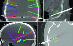 Generalisability and robustness tests. Our proposed algorithm using data from a different centre: a with smaller bolt heads, b contacts very close to each other, and c electrodes inserted deeply (pink electrode with insertion depth of 110 mm); Our data in SEEGA: d segmented contact positions (green fiducials) and implantation plan (pink fiducials). "We randomly chose 3 of our data sets to test the method proposed in [2, 16] and implemented in SEEGA 3D Slicer v4.6.2." |
Low-Cost Three-Dimensional Printed Phantom for Neuraxial Anesthesia Training: Development and Comparison to a Commercial Model
|
Publication: PLoS One. 2018 Jun 18;13(6):e0191664. PMID: 29912877 | PDF Authors: Mashari A, Montealegre-Gallegos M, Jeganathan J, Yeh L, Qua Hiansen J, Meineri M, Mahmood F, Matyal R. Institution: Department of Anesthesia and Pain Management, Toronto General Hospital, Toronto, Ontario, Canada. Abstract: METHODS: Anonymized CT DICOM data was segmented to create a 3D model of the lumbar spine. The 3D model was modified, placed inside a digitally designed housing unit and fabricated on a desktop 3D printer using polylactic acid (PLA) filament. The model was filled with an echogenic solution of gelatin with psyllium fiber. Twenty-two staff anesthesiologists performed a spinal and epidural on the 3D printed simulator and a commercially available Simulab phantom. Participants evaluated the tactile and ultrasound imaging fidelity of both phantoms via Likert-scale questionnaire. RESULTS: The 3D printed neuraxial phantom cost $13 to print and required 25 hours of non-supervised printing and 2 hours of assembly time. The 3D printed phantom was found to be less realistic to surface palpation than the Simulab phantom due to fragility of the silicone but had significantly better fidelity for loss of resistance, dural puncture and ultrasound imaging than the Simulab phantom. CONCLUSION: Low-cost neuraxial phantoms with fidelity comparable to commercial models can be produced using CT data and low-cost infrastructure consisting of FLOS software and desktop 3D printers. |
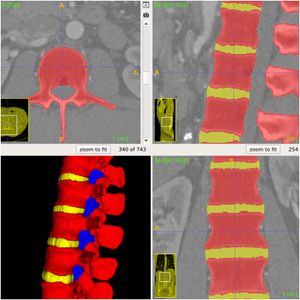 ITK-SNAP interface after segmentation of the spine with the automated active contour tool. Each colour represents a different layer-mask indicating different regions for the segmented 3D model. "We used the FLOS software ITK-SNAP and 3D Slicer for image segmentation". |
Improvement of Quality of 3D Printed Objects by Elimination of Microscopic Structural Defects in Fused Deposition Modeling
|
Publication: PLoS One. 2018 Jun 7;13(6):e0198370. PMID: 29879163 | PDF Authors: Gordeev EG, Galushko AS, Ananikov VP. Institution: N.D. Zelinsky Institute of Organic Chemistry, Russian Academy of Sciences, Moscow, Russia. Abstract: Additive manufacturing with fused deposition modeling (FDM) is currently optimized for a wide range of research and commercial applications. The major disadvantage of FDM-created products is their low quality and structural defects (porosity), which impose an obstacle to utilizing them in functional prototyping and direct digital manufacturing of objects intended to contact with gases and liquids. This article describes a simple and efficient approach for assessing the quality of 3D printed objects. Using this approach it was shown that the wall permeability of a printed object depends on its geometric shape and is gradually reduced in a following series: cylinder > cube > pyramid > sphere > cone. Filament feed rate, wall geometry and G-code-defined wall structure were found as primary parameters that influence the quality of 3D-printed products. Optimization of these parameters led to an overall increase in quality and improvement of sealing properties. It was demonstrated that high quality of 3D printed objects can be achieved using routinely available printers and standard filaments. "preparing the model for printing in the 3D Slicer program." |
Targeting HER2 Aberrations in Non-Small Cell Lung Cancer with Osimertinib
|
Publication: Clin Cancer Res. Clin Cancer Res. 2018 Jun 1;24(11):2594-604. PMID: 29298799 | PDF Authors: Liu S, Li S, Hai J, Wang X, Chen T, Quinn MM, Gao P, Zhang Y, Ji H, Cross D, Wong KK. Institution: Laboratory for Percutaneous Surgery, School of Computing, Queen's University, Kingston, ON, Canada. Abstract: Purpose: HER2 (or ERBB2) aberrations, including both amplification and mutations, have been classified as oncogenic drivers that contribute to 2-6 percent of lung adenocarcinomas. HER2 amplification is also an important mechanism for acquired resistance to EGFR tyrosine kinase inhibitors (TKIs). However, due to limited preclinical studies and clinical trials, currently there is still no available standard of care for lung cancer patients with HER2 aberrations. To fulfill the clinical need for targeting HER2 in non-small cell lung cancer (NSCLC) patients, we performed a comprehensive pre-clinical study to evaluate the efficacy of a third-generation TKI, osimertinib (AZD9291). Experimental Design:Three genetically modified mouse models (GEMMs) mimicking individual HER2 alterations in NSCLC were generated and osimertinib was tested for its efficacy against these HER2 aberrations in vivo. Results: Osimertinib treatment showed robust efficacy in HER2wt overexpression and EGFR del19/HER2 models but not in HER2 exon 20 insertion tumors. Interestingly, we further identified that combined treatment with osimertinib and the BET inhibitor JQ1 significantly increased the response rate in HER2-mutant NSCLC while JQ1 single treatment did not show efficacy. Conclusions: Overall, our data indicated robust anti-tumor efficacy of osimertinib against multiple HER2 aberrations in lung cancer, either as a single agent or in combination with JQ1. Our study provides a strong rationale for future clinical trials using osimertinib either alone or in combination with epigenetic drugs to target aberrant HER2 in NSCLC patients. "...Lung tumors were monitored by MRI and 3D Slicer was used to quantify the lung tumors." |
Plasma Membrane LAT Activation Precedes Vesicular Recruitment Defining Two Phases of Early T-cell Activation
|
Publication: Nat Commun. 2018 May 22;9(1):2013. PMID: 29789604 | PDF Authors: Balagopalan L, Yi J, Nguyen T, McIntire KM, Harned AS, Narayan K, Samelson LE. Institution: Laboratory of Cellular and Molecular Biology, Center for Cancer Research, National Cancer Institute, National Institutes of Health, Bethesda, MD, USA. Abstract: The relative importance of plasma membrane-localized LAT versus vesicular LAT for microcluster formation and T-cell receptor (TCR) activation is unclear. Here, we show the sequence of events in LAT microcluster formation and vesicle delivery, using lattice light sheet microscopy to image a T cell from the earliest point of activation. A kinetic lag occurs between LAT microcluster formation and vesicular pool recruitment to the synapse. Correlative 3D light and electron microscopy show an absence of vesicles at microclusters at early times, but an abundance of vesicles as activation proceeds. Using TIRF-SIM to look at the activated T-cell surface with high resolution, we capture directed vesicle movement between microclusters on microtubules. We propose a model in which cell surface LAT is recruited rapidly and phosphorylated at sites of T-cell activation, while the vesicular pool is subsequently recruited and dynamically interacts with microclusters. Funding:
|
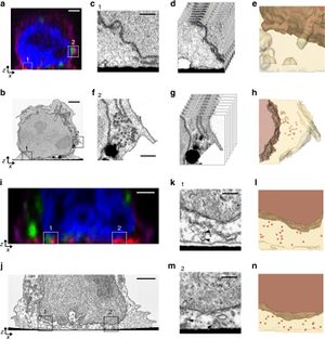 Correlative 3D light and FIB-SEM of activated T cells. Jurkat cells transfected with emerald-VAMP7 (green) were dropped onto stimulatory coverslips and fixed after 2 min a–h or 5 min i–n of activation and immunostained for pLAT (red), nucleus (blue), and plasma membrane (purple). a Images were collected of the whole cell activated for 2 min using confocal microscopy. "In the absence of correlative fiducial markers, “slabs” of volumetric data corresponding to the immune synapse from the confocal and FIB-SEM volumes were aligned using simple transforms in 3D Slicer." |
|
Publication: Surg Endosc. 2018 Jun;32(6):2713-20. PMID: 29214516 | PDF Authors: Wijsmuller AR, Romagnolo LGC, Agnus V, Giraudeau C, Melani AGF, Dallemagne B, Marescaux J. Institution: IRCAD/ EITS, Department of General, Digestive and Endocrine Surgery, Nouvel Hôpital Civil, University Hospital of Strasbourg, Strasbourg, France. Abstract: BACKGROUND: Stereotactic navigation could improve the quality of surgery for rectal cancer. Critical challenges related to soft tissue stereotactic pelvic navigation include the potential difference in patient anatomy between intraoperative lithotomy and preoperative supine position for imaging. The objective of this study was to determine the difference in patient anatomy, sacral tilt, and skin fiducial position between these different patient positions and to investigate the feasibility and optimal set-up for stereotactic pelvic navigation. METHODS: Four consecutive human anatomical specimens were submitted to repeated CT-scans in a supine and several degrees of lithotomy position. Patient anatomy, sacral tilt, and skin fiducial position were compared by means of an image computing platform. In two specimens, a 10-degree wedge was introduced to reduce the natural tilt of the sacrum during the shift from supine to lithotomy position. A simulation of laparoscopic and transanal surgical procedures was performed to assess the accuracy of the stereotactic navigation. RESULTS: An up-to-supracentimetric change in patient anatomy was noted between different patient positions. This observation was minimized through the application of a wedge. When switching from supine to another position, sacral retroversion occurred independent of the use of a wedge. There was considerable skin fiducial motion between different positions. Accurate stereotactic navigation was obtained with the least registration error (1.9 mm) when the position of the anatomical specimen was registered in a supine position with straight legs, without pneumoperitoneum, using a conventional CT-scan with an identical specimen positioning. CONCLUSION: The change in patient anatomy is small during the sacral tilt induced by positional changes when using a 10-degree wedge, allowing for an accurate stereotactic surgical navigation. This opens up new promising opportunities to increase the quality of surgery for rectal cancer cases where it is difficult or impossible to identify and dissect along the anatomical planes. "each skin fiducial were marked by using an image computing platform, 3D Slicer. |
 Markers were manually placed in the center of the skin fiducials in each CT-scan by using an image computing platform 3D Slicer (B). The other markers S1-6 (C) and P1-6 (D) were placed at the anatomical landmarks as depicted in Table 2. |
New Approach of Ultra-focal Brachytherapy for Low- and Intermediate-risk Prostate Cancer with Custom-linked I-125 Seeds: A feasibility Study of Optimal Dose Coverage
|
Publication: Brachytherapy. 2018 May - Jun;17(3):544-55. PMID: 29525514 Authors: Brun T, Bachaud JM, Graff-Cailleaud P, Malavaud B, Portalez D, Popotte C, Aziza R, Lusque A, Filleron T, Ken S. Institution: Institut Claudius Regaud, Institut Universitaire du Cancer de Toulouse - Oncopôle, Department of Engineering and Medical Physics, Toulouse, France. Abstract: PURPOSE: To present the feasibility study of optimal dose coverage in ultra-focal brachytherapy (UFB) with multiparametric MRI for low- and intermediate-risk prostate cancer. METHODS AND MATERIALS: UFB provisional dose plans for small target volumes (<7 cc) were calculated on a prostate training phantom to optimize the seeds number and strength. Clinical UFB consisted in a contour-based nonrigid registration (MRI/Ultrasound) to implant a fiducial marker at the location of the tumor focus. Dosimetry was performed with iodine-125 seeds and a prescribed dose of 160 Gy. On CT scans acquired at 1 month, dose coverage of 152 Gy to the ultra-focal gross tumor volume was evaluated. Registrations between magnetic resonance and CT scans were assessed on the first 8 patients with three software solutions: VariSeed, 3D Slicer, and Mirada, and quantitative evaluations of the registrations were performed. Impact of these registrations on the initial dose matrix was performed. RESULTS: Mean differences between simulated dose plans and extrapolated Bard nomogram for UFB volumes were 36.3% (26-56) for the total activity, 18.3% (10-30) for seed strength, and 22.5% (16-38) for number of seeds. Registration method implemented in Mirada performed significantly better than VariSeed and 3D Slicer (p = 0.0117 and p = 0.0357, respectively). For dose plan evaluation between Mirada and VariSeed, D100% (Gy) for ultra-focal gross tumor volume had a mean difference of 28.06 Gy, mean values being still above the objective of 152 Gy. D90% for the prostate had a mean difference of 1.17 Gy. For urethra and rectum, dose limits were far below the recommendations. CONCLUSIONS: This UFB study confirmed the possibility to treat with optimal dose coverage target volumes smaller than 7 cc. |
Evaluating the Association between Enlarged Perivascular Spaces and Disease Worsening in Multiple Sclerosis
|
Publication: J Neuroimaging. 2018 May;28(3):273-7. PMID: 29226505 Authors: Cavallari M, Egorova S, Healy BC, Palotai M, Prieto JC, Polgar-Turcsanyi M, Tauhid S, Anderson M, Glanz B, Chitnis T, Guttmann CRG. Institution: Partners Multiple Sclerosis Center, Brigham and Women's Hospital, Harvard Medical School, Boston, MA, USA. Abstract: BACKGROUND AND PURPOSE: Enlarged perivascular spaces (EPVSs) have been associated with relapses and brain atrophy in multiple sclerosis (MS). We investigated the association of EPVS with clinical and MRI features of disease worsening in a well-characterized cohort of relapsing-remitting MS patients prospectively followed for up to 10 years. METHODS: Baseline EPVSs were scored on 1.5T MRI in 30 converters to moderate-severe disability, and 30 nonconverters matched for baseline characteristics. RESULTS: EPVS scores were not significantly different between converters and nonconverters, nor associated with accrual of lesions or brain atrophy. CONCLUSIONS: Our preliminary findings from a relatively small study sample argue against a potential use of EPVS as early indicator of risk for disease worsening in relapsing-remitting MS patients in a clinical setting. Although the small sample size and clinical 1.5T MRI may have limited our ability to detect a significant effect, we provided estimates of the association of EPVS with clinical and MRI indicators of disease worsening in a well-characterized cohort of MS patients. "Outputs from the automated image analysis workflow were manually edited by a physician expert in image analysis (SE) in 3D Slicer" |
Clinical Evaluation of Semi-Automatic Open-Source Algorithmic Software Segmentation of the Mandibular Bone: Practical Feasibility and Assessment of a New Course of Action
|
Publication: PLoS One. 2018 May 10;13(5):e0196378. PMID: 29746490 | PDF Authors: Wallner J, Hochegger K, Chen X, Mischak I, Reinbacher K, Pau M, Zrnc T, Schwenzer-Zimmerer K, Zemann W, Schmalstieg D, Egger J. Institution: Department of Oral & Maxillofacial Surgery, Medical University of Graz, Graz, AT. Abstract: INTRODUCTION: Computer assisted technologies based on algorithmic software segmentation are an increasing topic of interest in complex surgical cases. However-due to functional instability, time consuming software processes, personnel resources or licensed-based financial costs many segmentation processes are often outsourced from clinical centers to third parties and the industry. Therefore, the aim of this trial was to assess the practical feasibility of an easy available, functional stable and licensed-free segmentation approach to be used in the clinical practice. MATERIAL AND METHODS: In this retrospective, randomized, controlled trail the accuracy and accordance of the open-source based segmentation algorithm GrowCut was assessed through the comparison to the manually generated ground truth of the same anatomy using 10 CT lower jaw data-sets from the clinical routine. Assessment parameters were the segmentation time, the volume, the voxel number, the Dice Score and the Hausdorff distance. RESULTS: Overall semi-automatic GrowCut segmentation times were about one minute. Mean Dice Score values of over 85% and Hausdorff Distances below 33.5 voxel could be achieved between the algorithmic GrowCut-based segmentations and the manual generated ground truth schemes. Statistical differences between the assessment parameters were not significant (p<0.05) and correlation coefficients were close to the value one (r > 0.94) for any of the comparison made between the two groups. DISCUSSION: Complete functional stable and time saving segmentations with high accuracy and high positive correlation could be performed by the presented interactive open-source based approach. In the cranio-maxillofacial complex the used method could represent an algorithmic alternative for image-based segmentation in the clinical practice for e.g. surgical treatment planning or visualization of postoperative results and offers several advantages. Due to an open-source basis the used method could be further developed by other groups or specialists. Systematic comparisons to other segmentation approaches or with a greater data amount are areas of future works. |
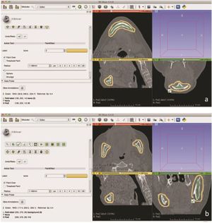 Axial Algorithmic (GrowCut) segmentation in 3D Slicer. (a) Fore- (green) and background (yellow) initialization of GrowCut in the lower jawbone in an axial, sagittal and coronal slice around the anterior mandible (symphysis / para-symphysis). (b) Slicer based algorithmic (GrowCut) segmentation: Fore- (green) and background (yellow) initialization of GrowCut in the lower jawbone in an axial, sagittal and coronal slice around parts of the mandible. |
Subthalamic Oscillatory Activity and Connectivity during Gait in Parkinson's Disease
|
Publication: Neuroimage Clin. 2018 May 3;19:396-405. PMID: 30035024 | PDF Authors: Hell F, Plate A, Mehrkens JH, Bötzel K. Institution: Department of Neurology, Ludwig-Maximilians University, Munich, Germany. Abstract: Local field potentials (LFP) of the subthalamic nucleus (STN) recorded during walking may provide clues for determining the function of the STN during gait and also, may be used as biomarker to steer adaptive brain stimulation devices. Here, we present LFP recordings from an implanted sensing neurostimulator (Medtronic Activa PC + S) during walking and rest with and without stimulation in 10 patients with Parkinson's disease and electrodes placed bilaterally in the STN. We also present recordings from two of these patients recorded with externalized leads. We analyzed changes in overall frequency power, bilateral connectivity, high beta frequency oscillatory characteristics and gait-cycle related oscillatory activity. We report that deep brain stimulation improves gait parameters. High beta frequency power (20-30 Hz) and bilateral oscillatory connectivity are reduced during gait, while the attenuation of high beta power is absent during stimulation. Oscillatory characteristics are affected in a similar way. We describe a reduction in overall high beta burst amplitude and burst lifetimes during gait as compared to rest off stimulation. Investigating gait cycle related oscillatory dynamics, we found that alpha, beta and gamma frequency power is modulated in time during gait, locked to the gait cycle. We argue that these changes are related to movement induced artifacts and that these issues have important implications for similar research. |
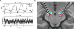 Shank rotation measurements, raw LFP recordings and electrode localization. A. Shank rotation velocity of two different goniometer sensors setups mounted at right and left leg of one patient during walking and raw LFP trace from a single STN recording with Activa PC + S sensing. B. Posterior dorsal view of DBS-electrode localizations in left and right STN. The motor region of the STN is depicted in dark red, the associative subregion in light blue and limbic subregion in yellow. The contact used for stimulation is colored in light red (see text for details). "MRT and CT were aligned manually using 3D Slicer software, co-registered using a two-stage linear registration (rigid followed by affine) as implemented in Advanced Normalization Tools" |
Complete Thoracolumbar Fracture-dislocation with Intact Neurologic Function: Explanation of a Novel Cord Saving Mechanism
|
Publication: J Spinal Cord Med. 2018 May;41(3):367-76. PMID: 28648115 | PDF Authors: Rahimizadeh A, Asgari N, Rahimizadeh A. Institution: Department of Neurosurgery, Pars Advanced and Minimally Invasive Medical Manners Research Center, Pars Hospital, Iran University of Medical Science, Tehran, Iran. Abstract: Background: The thoracolumbar junction from T11 to L2 is a common site of injury in which fracture and dislocations are the most prevalent ones occurring at this location. Fracture dislocation is defined as failure of all three columns of the spine with gross displacement. Considering the significant violence necessary to produce fracture dislocations, these injuries are often associated with major neural deficit, with the majority of casualties becoming paraplegic immediately. Preservation of neurological function following complete fracture dislocation is quite rare entity. Objective: To represent the possibility of existence of a preservation mechanism for functional integrity of cord despite spinal gross fracture dislocation by reproducing the injury on a plastic model and simulating a corresponding model using 3D Slicer software, detailed description the pathomechanism of neurologic sparing. Case Report: A 19-year-old female who sustained severe thoracolumbar fracture dislocation but with normal neurology is presented. Despite the severity of the condition, the diagnosis was initially missed due to associated vital injuries. Results: Combined posterior and anterior surgery resulted in optimal coronal and sagittal alignment, as well as proper stabilization without any complication. At 9-year follow-up, the patient was found to be doing well. Conclusion: The prognosis for complete recovery with preplanned surgical intervention in thoracolumbar injuries affecting all three columns but with normal neurologic function is promising based on images, plastic models and 3D simulated model based on digital images. |
Optimization of 3D Print Material for the Recreation of Patient-Specific Temporal Bone Models
|
Publication: Ann Otol Rhinol Laryngol. 2018 May;127(5):338-43. PMID: 29667491 Authors: Haffner M, Quinn A, Hsieh TY, Strong EB, Steele T. Institution: University of California, Davis, School of Medicine, Sacramento, CA, USA. Abstract: OBJECTIVE: Identify the 3D printed material that most accurately recreates the visual, tactile, and kinesthetic properties of human temporal bone Subjects and Methods: Fifteen study participants with an average of 3.6 years of postgraduate training and 56.5 temporal bone (TB) procedures participated. Each participant performed a mastoidectomy on human cadaveric TB and five 3D printed TBs of different materials. After drilling each unique material, participants completed surveys to assess each model's appearance and physical likeness on a Likert scale from 0 to 10 (0 = poorly representative, 10 = completely life-like). The 3D models were acquired by computed tomography (CT) imaging and segmented using 3D Slicer software. RESULTS: Polyethylene terephthalate (PETG) had the highest average survey response for haptic feedback (HF) and appearance, scoring 8.3 (SD = 1.7) and 7.6 (SD = 1.5), respectively. The remaining plastics scored as follows for HF and appearance: polylactic acid (PLA) averaged 7.4 and 7.6, acrylonitrile butadiene styrene (ABS) 7.1 and 7.2, polycarbonate (PC) 7.4 and 3.9, and nylon 5.6 and 6.7. CONCLUSION: A PETG 3D printed temporal bone models performed the best for realistic appearance and HF as compared with PLA, ABS, PC, and nylon. The PLA and ABS were reliable alternatives that also performed well with both measures. |
3-D Segmentation of Lung Nodules using Hybrid Level Sets
|
Publication: Comput Biol Med. 2018 May 1;96:214-26. PMID: 29631230 Authors: Shakir H, Rasool Khan TM, Rasheed H. Institution: Department of Electrical Engineering, Bahria University, 13-National Stadium Road, Karachi, Pakistan. Abstract: Lung nodule segmentation in CT images and its subsequent volume analysis can help determine the malignancy status of a lung nodule. While several efficient segmentation schemes have been proposed, only a few studies evaluated the segmentation's performance for large nodules. In this research, we contribute a semi-automatic system which is capable of performing robust 3-D segmentations on both small and large nodules with good accuracy. The target CT volume is de-noised with an anisotropic diffusion filter and a region of interest is selected around the target nodule on a reference slice. The proposed model performs nodule segmentation by incorporating a mean intensity based threshold in Geodesic Active Contour model in level sets. We also devise an adaptive technique using image intensity histogram to estimate the desired mean intensity of the nodule. The proposed system is validated on both lung nodules and phantoms collected from publicly available diverse databases. Quantitative and visual comparative analysis of the proposed work with the Chan-Vese algorithm and statistic active contour model of 3D Slicer platform is also presented. The resulting mean spatial overlap between segmented nodules and reference nodules is 0.855, the mean volume bias is 0.10±0.2 ml and the algorithm repeatability is 0.060 ml. The achieved results suggest that the proposed method can be used for volume estimations of small as well as large-sized nodules. |
Prediction of Outcome using Pretreatment 18F-FDG PET/CT and MRI Radiomics in Locally Advanced Cervical Cancer Treated with Chemoradiotherapy
|
Publication: Eur J Nucl Med Mol Imaging. 2018 May;45(5):768-86. PMID: 29222685 Authors: Lucia F, Visvikis D, Desseroit MC, Miranda O, Malhaire JP, Robin P, Pradier O, Hatt M, Schick U. Institution: Department of Radiation Oncology, University Hospital, Brest, France. Abstract: PURPOSE: The aim of this study is to determine if radiomics features from 18fluorodeoxyglucose (FDG) positron emission tomography/computed tomography (PET/CT) and magnetic resonance imaging (MRI) images could contribute to prognoses in cervical cancer. METHODS: One hundred and two patients (69 for training and 33 for testing) with locally advanced cervical cancer (LACC) receiving chemoradiotherapy (CRT) from 08/2010 to 12/2016 were enrolled in this study. 18F-FDG PET/CT and MRI examination [T1, T2, T1C, diffusion-weighted imaging (DWI)] were performed for each patient before CRT. Primary tumor volumes were delineated with the fuzzy locally adaptive Bayesian algorithm in the PET images and with 3D Slicer in the MRI images. Radiomics features (intensity, shape, and texture) were extracted and their prognostic value was compared with clinical parameters for recurrence-free and locoregional control. RESULTS: In the training cohort, median follow-up was 3.0 years (range, 0.43-6.56 years) and relapse occurred in 36% of patients. In univariate analysis, FIGO stage (I-II vs. III-IV) and metabolic response (complete vs. non-complete) were probably associated with outcome without reaching statistical significance, contrary to several radiomics features from both PET and MRI sequences. Multivariate analysis in training test identified Grey Level Non UniformityGLRLM in PET and EntropyGLCM in ADC maps from DWI MRI as independent prognostic factors. These had significantly higher prognostic power than clinical parameters, as evaluated in the testing cohort with accuracy of 94% for predicting recurrence and 100% for predicting lack of loco-regional control (versus ~50-60% for clinical parameters). CONCLUSIONS: In LACC treated with CRT, radiomics features such as EntropyGLCM and GLNUGLRLM from functional imaging DWI-MRI and PET, respectively, are independent predictors of recurrence and loco-regional control with significantly higher prognostic power than usual clinical parameters. Further research is warranted for their validation, which may justify more aggressive treatment in patients identified with high probability of recurrence. |
Smoking Duration Alone Provides Stronger Risk Estimates of Chronic Obstructive Pulmonary Disease than Pack-years
|
Publication: Thorax. 2018 May;73(5):414-21. PMID: 29326298 | PDF Authors: Bhatt SP, Kim YI, Harrington KF, Hokanson JE, Lutz SM, Cho MH, DeMeo DL, Wells JM, Make BJ, Rennard SI, Washko GR, Foreman MG, Tashkin DP, Wise RA, Dransfield MT, Bailey WC; COPDGene Investigators. Institution: Division of Pulmonary, Allergy and Critical Care Medicine, University of Alabama at Birmingham, Birmingham, AL, USA. Abstract: Cigarette smoking is the strongest risk factor for COPD. Smoking burden is frequently measured in pack-years, but the relative contribution of cigarettes smoked per day versus duration towards the development of structural lung disease, airflow obstruction and functional outcomes is not known. METHODS: We analyzed cross-sectional data from a large multi-centre cohort (COPDGene) of current and former smokers. Primary outcome was airflow obstruction (FEV1/FVC); secondary outcomes included five additional measures of disease: FEV1, CT emphysema, CT gas trapping, functional capacity (6 min walk distance, 6MWD) and respiratory morbidity (St George's Respiratory Questionnaire, SGRQ). Generalized linear models were estimated to compare the relative contribution of each smoking variable with the outcomes, after adjustment for age, race, sex, body mass index, CT scanner, centre, age of smoking onset and current smoking status. We also estimated adjusted means of each outcome by categories of pack-years and combined groups of categorized smoking duration and cigarettes/day, and estimated linear trends of adjusted means for each outcome by categorized cigarettes/day, smoking duration and pack-years. RESULTS: 10 187 subjects were included. For FEV1/FVC, standardized beta coefficient for smoking duration was greater than for cigarettes/day and pack-years (P<0.001). After categorization, there was a linear increase in adjusted means FEV1/FVC with increase in pack-years (regression coefficient β=-0.023±SE0.003; P=0.003) and duration over all ranges of smoking cigarettes/day (β=-0.041±0.004; P<0.001) but a relatively flat slope for cigarettes/day across all ranges of smoking duration (β=-0.009±0.0.009; P=0.34). Strength of association of duration was similarly greater than pack-years for emphysema, gas trapping, FEV1, 6MWD and SGRQ. CONCLUSION: Smoking duration alone provides stronger risk estimates of COPD than the composite index of pack-years. "... After segmentation and extrusion of the large and medium-sized airways, emphysema was quantified as the percentage of lung volume at TLC with attenuation <−950 Hounsfield units (HU) by density mask analyses using 3D Slicer software." |
Using 3DSlicer, Z-Brush, and Slic3r to Turn CAT Scans Into Kidney 3D-Prints
|
Publication: Adafruit, May 24, 2018. Authors: Andrew Krill Read the article here |
Development of White Matter Microstructure in Relation to Verbal and Visuospatial Working Memory-A Longitudinal Study
|
Publication: PLoS One. 2018 Apr 24;13(4):e0195540. PMID: 29689058 | PDF Authors: Krogsrud SK, Fjell AM, Tamnes CK, Grydeland H, Due-Tønnessen P, Bjørnerud A, Sampaio-Baptista C, Andersson J, Johansen-Berg H, Walhovd KB. Institution: Research Group for Lifespan Changes in Brain and Cognition, Department of Psychology, University of Oslo, Oslo, Norway. Abstract: Working memory capacity is pivotal for a broad specter of cognitive tasks and develops throughout childhood. This must in part rely on development of neural connections and white matter microstructure maturation, but there is scarce knowledge of specific relations between this and different aspects of working memory. Diffusion tensor imaging (DTI) enables us to study development of brain white matter microstructure. In a longitudinal DTI study of 148 healthy children between 4 and 11 years scanned twice with an on average 1.6 years interval, we characterized change in fractional anisotropy (FA), mean (MD), radial (RD) and axial diffusivity (AD) in 10 major white matter tracts hypothesized to be of importance for working memory. The results showed relationships between change in several tracts and change in visuospatial working memory. Specifically, improvement in visuospatial working memory capacity was significantly associated with decreased MD, RD and AD in inferior longitudinal fasciculus (ILF), inferior fronto-occipital fasciculus (IFOF) and uncinate fasciculus (UF) in the right hemisphere, as well as forceps major (FMaj). No significant relationships were found between change in DTI metrics and change in verbal working memory capacity. These findings yield new knowledge about brain development and corresponding working memory improvements in childhood. |
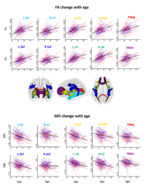 FA and MD in specific tracts with age. Spaghetti plots of individual participant change in FA and MD in specific tracts with age (years). Females are plotted in red and males in blue. For each measure, an assumption-free general additive mixed model as a function of age was fitted to accurately describe change across the age range. Three-dimensional renderings illustrate ten atlas-based probabilistic tracts from the Mori atlas in anterior, left, and dorsal views, displayed on a semitransparent template brain. The color-coded titles for each scatterplot represent the color of each specific white matter tract. Color codes refer to: Light blue: Inferior longitudinal fasciculus (ILF), Yellow: Inferior fronto-occipital fasciculus (IFOF), Red: Forceps major (FMaj), Blue: Superior longitudinal fasciculus (SLF), Green: Uncinate fasciculus (UF), and Purple: Forceps minor (FMin). The 3D figures were made by the use of 3D Slicer. L = left and R = right. |
Detailed T1-Weighted Profiles from the Human Cortex Measured in Vivo at 3 Tesla MRI
|
Publication: Neuroinformatics. 2018 Apr;16(2):181-96. PMID: 29352389 | PDF Authors: Ferguson B, Petridou N, Fracasso A, van den Heuvel MP, Brouwer RM, Hulshoff Pol HE, Kahn RS, Mandl RCW. Institution: Rudolf Magnus Brain Center, Brain Division, Department of Psychiatry, University Medical Center Utrecht, Utrecht University, The Netherlands. Abstract: Studies into cortical thickness in psychiatric diseases based on T1-weighted MRI frequently report on aberrations in the cerebral cortex. Due to limitations in image resolution for studies conducted at conventional MRI field strengths (e.g. 3 Tesla (T)) this information cannot be used to establish which of the cortical layers may be implicated. Here we propose a new analysis method that computes one high-resolution average cortical profile per brain region extracting myeloarchitectural information from T1-weighted MRI scans that are routinely acquired at a conventional field strength. To assess this new method, we acquired standard T1-weighted scans at 3 T and compared them with state-of-the-art ultra-high resolution T1-weighted scans optimised for intracortical myelin contrast acquired at 7 T. Average cortical profiles were computed for seven different brain regions. Besides a qualitative comparison between the 3 T scans, 7 T scans, and results from literature, we tested if the results from dynamic time warping-based clustering are similar for the cortical profiles computed from 7 T and 3 T data. In addition, we quantitatively compared cortical profiles computed for V1, V2 and V7 for both 7 T and 3 T data using a priori information on their relative myelin concentration. Although qualitative comparisons show that at an individual level average profiles computed for 7 T have more pronounced features than 3 T profiles the results from the quantitative analyses suggest that average cortical profiles computed from T1-weighted scans acquired at 3 T indeed contain myeloarchitectural information similar to profiles computed from the scans acquired at 7 T. The proposed method therefore provides a step forward to study cortical myeloarchitecture in vivo at conventional magnetic field strength both in health and disease. Funding:
|
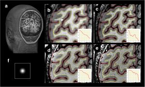 Result of deconvolution of 3 T and 7 T scans, coronal slice. Effect of deconvolution shown in a coronal section of the occipital lobe for one participant for the area of interest displayed in the white rectangle in (a). The four panels on the right indicate the area of interest in an original (a) and deconvoluted (b) 3 T T1w 0.8 mm isotropic scan and in an original (d) and deconvoluted (e) 7 T myelin-sensitive T1w 0.5 mm isotropic scan. Freesurfer white (red) and pial (yellow) surfaces are overlaid. Insets display signal intensity sampled along displayed orange ruler (marked by the white arrow), with 3D Slicer 4.4.0, which is 4.2 mm in length and runs from white matter to CSF. Please note that this sample is drawn in a 2D projection and is not necessarily perpendicular to the cortical mantle. (f) depicts the PSF used |
Estimating Shape Correspondence for Populations of Objects with Complex Topology
|
Publication: Proc IEEE Int Symp Biomed Imaging. 2018 Apr;2018:1010-3. PMID: 29973974 | PDF Authors: Fishbaugh J, Pascal L, Fischer L, Nguyen T, Boen C, Goncalves J, Gerig G, Paniagua B. Institution: NYU Tandon School of Engineering, NY, USA. Abstract: Statistical shape analysis captures the geometric properties of a given set of shapes, obtained from medical images, by means of statistical methods. Orthognathic surgery is a type of craniofacial surgery that is aimed at correcting severe skeletal deformities in the mandible and maxilla. Methods assuming spherical topology cannot represent the class of anatomical structures exhibiting complex geometries and topologies, including the mandible. In this paper we propose methodology based on non-rigid deformations of 3D geometries to be applied to objects with thin, complex structures. We are able to accurately and quantitatively characterize bone healing at the osteotomy site as well as condylar remodeling for three orthognathic surgery cases, demonstrating the effectiveness of the proposed methodology. Funding:
|
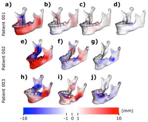 Signed corresponding distance computed between a-e-h) T1 and T2 shapes, displayed on T1, b-f-i) T2 and T3, displayed on T2, c-f-i) T3 and T4, displayed on T3 and d) T4 and T5, displayed on T4. "Measurements were made using ModelToModelDistance [14], a surface to surface distance plugin in 3D Slicer." |
Development of a Hybrid Computational/Experimental Framework for Evaluation of Damage Mechanisms of a Linked Semiconstrained Total Elbow System
|
Publication: J Shoulder Elbow Surg. 2018 Apr;27(4):614-23. PMID: 29305101 Authors: Sharifi Kia D, Willing R. Institution: Biomechanics and Orthopedic Design Laboratory, Department of Mechanical Engineering, State University of New York at Binghamton, Binghamton, NY, USA. Abstract: Background: Long-term durability of total elbow arthroplasty (TEA) is a concern, and bearing wear or excessive deformations may necessitate early revision. The current study used experimental wear testing and computational finite element modeling to develop a hybrid computational and experimental framework for the evaluation of TEA damage mechanisms. Methods: Three Coonrad-Morrey (Zimmer-Biomet Inc., Warsaw, IN, USA) TEA implants were used for experimental wear testing for 200,000 cycles. Gravimetric measurements were performed before and after the tests to assess the weight change caused by wear. A finite element model of the implant was also developed to analyze ultrahigh-molecular-weight polyethylene (UHMWPE) damage. Results: High localized contact pressures caused visible creep and plastic flow, deforming bushings and creating unintended UHMWPE-on-UHMWPE contact surfaces where considerably high wear rates were observed. Average experimentally measured vs. model-predicted wear was 9.5 ± 1.0 vs. 14.1 mg for the of the medial bushing, 8.5 ± 1.0 vs. 13.9 mg for the lateral humeral bushing, and 34.1 ± 0.7 vs. 36.9 mg for the ulnar bushings, respectively. Model predicted contact stresses on the surfaces of bushings were substantially higher than the yield limit of conventional UHMWPE (87 MPa for the humeral bushings and 83 MPa for the ulnar bushing). Conclusions: Our study discovered that unintended wear at UHMWPE-UHMWPE contact surfaces, "fed" by excessive plastic flow may, in fact, be of more concern than wear that occurs at the intended metal-UHMWPE contact interfaces. Furthermore, formation of high localized contact stresses much above the yield limit of UHMWPE is another likely contributor to bushing failure for this implant. "...files were imported into 3D Slicer for 3-dimensional (3D) model reconstruction." |
Simulation of the Human Airways using Virtual Topology Tools and Meshing Optimization
|
Publication: Biomech Model Mechanobiol. 2018 Apr;17(2):465-77. PMID: 29105007 Authors: Fernández-Tena A, Marcos AC, Agujetas R, Ferrera C. Institution: Central University Hospital of Asturias, Oviedo, Spain. Abstract: A method is proposed to improve the quality of the three-dimensional airway geometric models using a commercial software, checking the number of elements, meshing time, and aspect ratio and skewness parameters. The use of real and virtual topologies combined with patch-conforming and patch-independent meshing algorithms results in four different models being the best solution the combination of virtual topology and patch-independent algorithm, due to an excellent aspect ratio and skewness of the elements, and minimum meshing time. The result is a reduction in the computational time required for both meshing and simulation due to a smaller number of cells. The use of virtual topologies combined with patch-independent meshing algorithms could be extended in bioengineering because the geometries handling is similar to this case. The method is applied to a healthy person using their computed tomography images. The resulting numerical models are able to simulate correctly a forced spirometry. "As all human tissues are classified under the Hounsfield scale, the 3D Slicer software was used to group similar grey values, identifying the threshold between the different tissues and extracting the human airways." |
Planning of Skull Reconstruction Based on a Statistical Shape Model Combined with Geometric Morphometrics
|
Publication: Int J Comput Assist Radiol Surg. 2018 Apr;13(4):519-29. PMID: 29080945 Authors: Fuessinger MA, Schwarz S, Cornelius CP, Metzger MC, Ellis E 3rd, Probst F, Semper-Hogg W, Gass M, Schlager S. Institution: Department of Oral and Maxillofacial Surgery, Albert-Ludwigs University, Freiburg, Germany. Abstract: Purpose: Virtual reconstruction of large cranial defects is still a challenging task. The current reconstruction procedures depend on the surgeon's experience and skills in planning the reconstruction based on mirroring and manual adaptation. The aim of this study is to propose and evaluate a computer-based approach employing a statistical shape model (SSM) of the cranial vault. Methods: An SSM was created based on 131 CT scans of pathologically unaffected adult crania. After segmentation, the resulting surface mesh of one patient was established as template and subsequently registered to the entire sample. using the registered surface meshes, an SSM was generated capturing the shape variability of the cranial vault. The knowledge about this shape variation in healthy patients was used to estimate the missing parts. The accuracy of the reconstruction was evaluated by using 31 CT scans not included in the SSM. Both unilateral and bilateral bony defects were created on each skull. The reconstruction was performed using the current gold standard of mirroring the intact to the affected side, and the result was compared to the outcome of our proposed SSM-driven method. The accuracy of the reconstruction was determined by calculating the distances to the corresponding parts on the intact skull. Results: While unilateral defects could be reconstructed with both methods, the reconstruction of bilateral defects was, for obvious reasons, only possible employing the SSM-based method. Comparing all groups, the analysis shows a significantly higher precision of the SSM group, with a mean error of 0.47 mm compared to the mirroring group which exhibited a mean error of 1.13 mm. Reconstructions of bilateral defects yielded only slightly higher estimation errors than those of unilateral defects. Conclusion: The presented computer-based approach using SSM is a precise and simple tool in the field of computer-assisted surgery. It helps to reconstruct large-size defects of the skull considering the natural asymmetry of the cranium and is not limited to unilateral defects. "Segmentation was done with 3D Slicer". |
Contribution of 3D Printing to Mandibular Reconstruction after Cancer
|
Publication: Eur Ann Otorhinolaryngol Head Neck Dis. 2018 Apr;135(2):133-6. PMID: 29100719 Authors: Dupret-Bories A, Vergez S, Meresse T, Brouillet F, Bertrand G. Institution: Chirurgie ORL et cervico-faciale, Institut Universitaire du Cancer Toulouse Oncopole, Institut Claudius-Regaud, Toulouse, France. Abstract: Three-dimensional (3D) printing is booming in the medical field. This technology increases the possibilities of personalized treatment for patients, while lowering manufacturing costs. To facilitate mandibular reconstruction with fibula free flap, some companies propose cutting guides obtained by CT-guided moulding. However, these guides are prohibitively expensive (€2,000 to €6,000). Based on a partnership with the CNRS, engineering students and a biomedical company, the authors have developed cutting guides and 3D-printed mandible templates, deliverable in 7days and at a lower cost. The novelty of this project is the speed of product development at a significantly lower price. In this technical note, the authors describe the logistic chain of production of mandible templates and cutting guides, as well as the results obtained. The goal is to allow access to this technology to all patients in the near future. "In order to demonstrate the feasibility and potential cost reduction, we preferred to use a free access software suite (3D Slicer, Blender, 3D Builder)" |
Optimizing Image Quantification for 177Lu SPECT/CT Based on a 3D Printed 2-Compartment Kidney Phantom
|
Publication: J Nucl Med. 2018 Apr;59(4):616-24. PMID: 29097409 | PDF Authors: Tran-Gia J, Lassmann M. Institution: University of Würzburg, Germany. Abstract: Aims: The aim of this work was to find an optimal setup for activity determination of 177Lu-based single photon emission computed tomography (SPECT) / computed tomography (CT) imaging reconstructed with two commercially available reconstructions (xSPECT Quant and Flash3D, Siemens Healthcare). For this purpose, 3D printed phantoms of different geometries were manufactured, different partial volume correction (PVC) methods were applied, and the accuracy of the activity determination was evaluated. Methods: A 2-compartment kidney phantom (70% cortical and 30% medullary compartment), a sphere, and an ellipsoid of equal volumes were 3D printed, filled with 177Lu, and scanned with a SPECT/CT system. Reconstructions were performed with xSPECT and Flash3D. Different PVC methods were applied to find an optimal quantification setup: 1) Geometry-specific recovery coefficient based on the 3D printing model. 2) Geometry-specific recovery coefficient based on the low-dose CT. 3) Enlarged volume-of-interest (VOI) including spilled-out counts. 4) Activity concentration in the peak milliliter applied to the entire CT-based volume. 5) Fixed threshold of 42% of the maximum in a large volume containing the object-of-interest. Additionally, the influence of post-reconstruction Gaussian filtering was investigated. Results: While the recovery coefficients of sphere and ellipsoid only differed by 0.7%, a difference of 31.7% was observed between the sphere and renal cortex phantoms . Without post-filtering, the model-based recovery coefficients (methods 1 and 2) resulted in the best accuracies (xSPECT: 1.5%, Flash3D: 10.3%), followed by the enlarged volume (xSPECT: 8.5%, Flash3D: 13.0%). The peak-milliliter method showed large errors only for sphere and ellipsoid (xSPECT: 23.4%, Flash3D: 21.6%). Applying a 42%-threshold led to the largest quantification errors (xSPECT: 32.3%, Flash3D: 46.7%). After post-filtering, a general increase of the errors was observed. Conclusion: In this work, 3D printing was used as prototyping technique for a geometry-specific investigation of SPECT/CT reconstruction parameters and PVC methods. An optimal setup for activity determination was found to be an unsmoothed SPECT/CT reconstruction in combination with a recovery coefficient calculated based on the low-dose CT. The difference between spherical and renal recovery coefficients suggests that the typically applied volume-dependent but only sphere-based recovery coefficient lookup tables should be replaced by a more geometry-specific alternative. "The highly-resolved mask as well as the filling volume were extracted from the low-dose CT using 3D Slicer." |
Endocardial Infarct Scar Recognition by Myocardial Electrical Impedance is not Influenced by Changes in Cardiac Activation Sequence
|
Publication: Heart Rhythm. 2018 Apr;15(4):589-96. PMID: 29197656 Authors: Amorós-Figueras G, Jorge E, Alonso-Martin C, Traver D, Ballesta M, Bragós R, Rosell-Ferrer J, Cinca J. Institution: Department of Cardiology, Hospital de la Santa Creu, Autonomous University of Barcelona, Barcelona, Spain. Abstract: Background: Measurement of myocardial electrical impedance can allow recognition of infarct scar and is theoretically not influenced by changes in cardiac activation sequence, but this is not known. Objectives: The objectives of this study were to evaluate the ability of endocardial electrical impedance measurements to recognize areas of infarct scar and to assess the stability of the impedance data under changes in cardiac activation sequence. Methods: One-month-old myocardial infarction confirmed by cardiac magnetic resonance imaging was induced in 5 pigs submitted to coronary artery catheter balloon occlusion. Electroanatomic data and local electrical impedance (magnitude, phase angle, and amplitude of the systolic-diastolic impedance curve) were recorded at multiple endocardial sites in sinus rhythm and during right ventricular pacing. By merging the cardiac magnetic resonance and electroanatomic data, we classified each impedance measurement site either as healthy (bipolar amplitude ≥1.5 mV and maximum pixel intensity <40%) or scar (bipolar amplitude <1.5 mV and maximum pixel intensity ≥40%). Results: A total of 137 endocardial sites were studied. Compared to healthy tissue, areas of infarct scar showed 37.4% reduction in impedance magnitude (P < .001) and 21.5% decrease in phase angle (P < .001). The best predictive ability to detect infarct scar was achieved by the combination of the 4 impedance parameters (area under the receiver operating characteristic curve 0.96; 95% confidence interval 0.92-1.00). In contrast to voltage mapping, right ventricular pacing did not significantly modify the impedance data. Conclusion: Endocardial catheter measurement of electrical impedance can identify infarct scar regions, and in contrast to voltage mapping, the impedance data are not affected by changes in cardiac activation sequence. "Processing of the LGE-CMR data was performed using the 3D Slicer software." |
Regional Hippocampal Vulnerability in Early Multiple Sclerosis: Dynamic Pathological Spreading from Dentate Gyrus to CA1
|
Publication: Hum Brain Mapp. 2018 Apr;39(4):1814-24. PMID: 29331060 | PDF Authors: Planche V, Koubiyr I, Romero JE, Manjon JV, Coupé P, Deloire M, Dousset V, Brochet B, Ruet A, Tourdias T. Institution: Universiry of Bordeaux, Bordeaux, France. Abstract: BACKGROUND: Whether hippocampal subfields are differentially vulnerable at the earliest stages of multiple sclerosis (MS) and how this impacts memory performance is a current topic of debate. METHOD: We prospectively included 56 persons with clinically isolated syndrome (CIS) suggestive of MS in a 1-year longitudinal study, together with 55 matched healthy controls at baseline. Participants were tested for memory performance and scanned with 3 T MRI to assess the volume of 5 distinct hippocampal subfields using automatic segmentation techniques. RESULTS: At baseline, CA4/dentate gyrus was the only hippocampal subfield with a volume significantly smaller than controls (p < .01). After one year, CA4/dentate gyrus atrophy worsened (-6.4%, p < .0001) and significant CA1 atrophy appeared (both in the stratum-pyramidale and the stratum radiatum-lacunosum-moleculare, -5.6%, p < .001 and -6.2%, p < .01, respectively). CA4/dentate gyrus volume at baseline predicted CA1 volume one year after CIS (R2 = 0.44 to 0.47, p < .001, with age, T2 lesion-load, and global brain atrophy as covariates). The volume of CA4/dentate gyrus at baseline was associated with MS diagnosis during follow-up, independently of T2-lesion load and demographic variables (p < .05). Whereas CA4/dentate gyrus volume was not correlated with memory scores at baseline, CA1 atrophy was an independent correlate of episodic verbal memory performance one year after CIS (ß = 0.87, p < .05). CONCLUSION: The hippocampal degenerative process spread from dentate gyrus to CA1 at the earliest stage of MS. This dynamic vulnerability is associated with MS diagnosis after CIS and will ultimately impact hippocampal-dependent memory performance. "Binary maps of lesions were reviewed and corrected manually by two blinded experts (MR engineer and neurologist), using 3D Slicer 4.4.0." |
Effects of Total Saponins from Trillium Tschonoskii Rhizome on Grey and White Matter Injury Evaluated by Quantitative Multiparametric MRI in a Rat Model of Ischemic Stroke
|
Publication: J Ethnopharmacol. 2018 Apr 6;215:199-209. PMID: 29309860 Authors: Li M, Ouyang J, Zhang Y, Cheng BCY, Zhan Y, Yang L, Zou H, Zhao H. Institution: School of Traditional Chinese Medicine, Capital Medical University, Beijing, China. Abstract: Ethnopharmacological Relevance: Trillium tschonoskii rhizome (TTR), a medicinal herb, has been traditionally used to treat traumatic brain injury and headache in China. Although the potential neuroprotective efficacy of TTR has gained increasing interest, the pharmacological mechanism remains unclear. Steroid saponins are the main bioactive components of the herb. Aim of Study: To investigate the protective and repair-promoting effects of the total saponins from TTR (TSTT) on grey and white matter damages in a rat model of middle cerebral artery occlusion (MCAO) using magnetic resonance imaging (MRI) assay. Materials and Methods: Ischemic stroke was induced by MCAO. TSTT and Ginaton (positive control) were administered orally to rats 6h after stroke and daily thereafter. After 15 days of treatment, the survival rate of each group was calculated. We then conducted neurological deficit scores and beam walking test to access the neurological function after ischemic stroke. Subsequently, T2-weighted imaging (T2WI) and T2 relaxometry mapping were performed to measure infarct volume and grey and white matter integrity, respectively. Moreover, diffusion tensor imaging (DTI) was carried out to evaluate the grey and white matter microstructural damage. Additionally, arterial spin labelling (ASL) - cerebral blood flow (CBF) and magnetic resonance angiography (MRA) images provided dynamic information about vascular hemodynamic dysfunction after ischemic stroke. Finally, haematoxylin and eosin (HE) staining was carried out to evaluate the stroke-induced pathological changes in the brain. Results: The survival rate and neurological behavioural outcomes (Bederson scores and beam walking tests) were markedly ameliorated by TSTT (65mg/kg) treatment within 15 days after ischemic stroke. Moreover, T2WI and T2 relaxometry mapping showed that TSTT (65mg/kg) significantly reduced infarct volume and attenuated grey and white matter injury, respectively, which was confirmed by histopathological evaluation of brain tissue. The results obtained from DTI showed that TSTT (65mg/kg) not only significantly alleviated axonal damage and demyelination, but also promoted axonal remodelling and re-myelination. In addition, TSTT treatment also enhanced vascular signal density and increased CBF in rats after MCAO. Conclusion: Our results suggested the potential protective and repair-promoting effects of TSTT on grey and white matter from damage induced by ischemia. This study provides a modern pharmacological basis for the application of TSTT in managing ischemic stroke. "To determine the orientation and integrity of fibre systems, fibre tractography was conducted using 3D Slicer software." |
Optimized Programming Algorithm for Cylindrical and Directional Deep Brain Stimulation Electrodes
|
Publication: J Neural Eng. 2018 Apr;15(2):026005. PMID: 29235446 | PDF Authors: Anderson DN, Osting B, Vorwerk J, Dorval AC, Butson CR. Institution: Bioengineering, University of Utah, Salt Lake City, UT, USA. Abstract: Deep brain stimulation (DBS) is a growing treatment option for movement and psychiatric disorders. As DBS technology moves toward directional leads with increased numbers of smaller electrode contacts, trial-and-error methods of manual DBS programming are becoming too time-consuming for clinical feasibility. We propose an algorithm to automate DBS programming in near real-time for a wide range of DBS lead designs. Approach: Magnetic resonance imaging and diffusion tensor imaging are used to build finite element models that include anisotropic conductivity. The algorithm maximizes activation of target tissue and utilizes the Hessian matrix of the electric potential to approximate activation of neurons in all directions. We demonstrate our algorithm's ability in an example programming case that targets the sub-thalamic nucleus (STN) for the treatment of Parkinson's disease for three lead designs: the Medtronic 3389 (four cylindrical contacts), the direct STNAcute (two cylindrical contacts, six directional contacts), and the Medtronic-Sapiens lead (40 directional contacts). Main Results: The optimization algorithm returns patient-specific contact configurations in near real-time - less than ten seconds for even the most complex leads. When the lead was placed centrally in the target STN, the directional leads were able to activate over 50% of the region whereas the Medtronic 3389 could only activate 40%. When the lead was placed 2 mm lateral to the target, the directional leads performed as well as they did in the central position, but the Medtronic 3389 only activated 2.9% of the STN. Significance: This DBS programming algorithm can be applied to cylindrical electrodes as well as novel directional leads that are too complex with modern technology to be manually programmed. This algorithm may reduce clinical programming time and encourage the use of directional leads since they activate a larger volume of the target area than cylindrical electrodes in central and off-target lead placements. "...used a deterministic streamline algorithm in 3D Slicer software package." |
Temporomandibular Joint Regeneration: Proposal of a Novel Treatment for Condylar Resorption after Orthognathic Surgery using Transplantation of Autologous Nasal Septum Chondrocytes, and the First Human Case Report
|
Publication: Stem Cell Res Ther. 2018 Apr 7;9(1):94. PMID: 29625584 | PDF Authors: de Souza Tesch R, Takamori ER, Menezes K, Carias RBV, Dutra CLM, de Freitas Aguiar M, Torraca TSS, Senegaglia AC, Rebelatto CLK, Daga DR, Brofman PRS, Borojevic R. Institution: Centro de Medicina Regenerativa, Faculdade de Medicina de Petrópolis - FASE, Petrópolis, Brazil. Abstract: BACKGROUND: Upon orthognathic mandibular advancement surgery the adjacent soft tissues can displace the distal bone segment and increase the load on the temporomandibular joint causing loss of its integrity. Remodeling of the condyle and temporal fossa with destruction of condylar cartilage and subchondral bone leads to postsurgical condylar resorption, with arthralgia and functional limitations. Patients with severe lesions are refractory to conservative treatments, leading to more invasive therapies that range from simple arthrocentesis to open surgery and prosthesis. Although aggressive and with a high risk for the patient, surgical invasive treatments are not always efficient in managing the degenerative lesions. METHODS: We propose a regenerative medicine approach using in-vitro expanded autologous cells from nasal septum applied to the first proof-of-concept patient. After the required quality controls, the cells were injected into each joint by arthrocentesis. Results were monitored by functional assays and image analysis using computed tomography. RESULTS: The cell injection fully reverted the condylar resorption, leading to functional and structural regeneration after 6 months. Computed tomography images showed new cortical bone formation filling the former cavity space, and a partial recovery of condylar and temporal bones. The superposition of the condyle models showed the regeneration of the bone defect, reconstructing the condyle original form. CONCLUSIONS: We propose a new treatment of condylar resorption subsequent to orthognathic surgery, presently treated only by alloplastic total joint replacement. We propose an intra-articular injection of autologous in-vitro expanded cells from the nasal septum. The proof-of-concept treatment of a selected patient that had no alternative therapeutic proposal has given promising results, reaching full regeneration of both the condylar cartilage and bone at 6 months after the therapy, which was fully maintained after 1 year. This first case is being followed by inclusion of new patients with a similar pathological profile to complete an ongoing stage I/II study. |
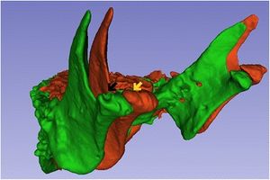 CT image three-dimensional reconstruction, being green before and red after the cell therapy injection. Note the filling of the bony defect in the right TMJ (black and yellow arrows). "After the segmentation, the next step of image analysis consisted of registering the scans and their respective three-dimensional volumetric models in a common coordinate system using a target region as the reference. This was done with 3D Slicer v.3.1 software." |
Validation of MRI to TRUS Registration for High-dose-rate Prostate Brachytherapy
|
Publication: Brachytherapy. 2018 Mar - Apr;17(2):283-90. PMID: 29331575 Authors: Poulin E, Boudam K, Pinter C, Kadoury S, Lasso A, Fichtinger G, Ménard C. Institution: Laboratory for Percutaneous Surgery, School of Computing, Queen's University, Kingston, Canada. Abstract: PURPOSE: The objective of this study was to develop and validate an open-source module for MRI to transrectal ultrasound (TRUS) registration to support tumor-targeted prostate brachytherapy. METHODS AND MATERIALS: In this study, 15 patients with prostate cancer lesions visible on multiparametric MRI were selected for the validation. T2-weighted images with 1-mm isotropic voxel size and diffusion weighted images were acquired on a 1.5T Siemens imager. Three-dimensional (3D) TRUS images with 0.5-mm slice thickness were acquired. The investigated registration module was incorporated in the open-source 3D Slicer platform, which can compute rigid and deformable transformations. An extension of 3D Slicer, SlicerRT, allows import of and export to DICOM-RT formats. For validation, similarity indices, prostate volumes, and centroid positions were determined in addition to registration errors for common 3D points identified by an experienced radiation oncologist. RESULTS: The average time to compute the registration was 35 ± 3 s. For the rigid and deformable registration, respectively, Dice similarity coefficients were 0.87 ± 0.05 and 0.93 ± 0.01 while the 95% Hausdorff distances were 4.2 ± 1.0 and 2.2 ± 0.3 mm. MRI volumes obtained after the rigid and deformable registration were not statistically different (p > 0.05) from reference TRUS volumes. For the rigid and deformable registration, respectively, 3D distance errors between reference and registered centroid positions were 2.1 ± 1.0 and 0.4 ± 0.1 mm while registration errors between common points were 3.5 ± 3.2 and 2.3 ± 1.1 mm. Deformable registration was found significantly better (p < 0.05) than rigid registration for all parameters. CONCLUSIONS: An open-source MRI to TRUS registration platform was validated for integration in the brachytherapy workflow. |
Commissioning and Validation of Commercial Deformable Image Registration Software for Adaptive Contouring
|
Publication: Phys Med. 2018 Mar;47:1-8. PMID: 29609810 Authors: Jamema SV, Phurailatpam R, Paul SN, Joshi K, Deshpande DD. Institution: Department of Radiation Oncology, Advanced Centre for Treatment Research and Education in Cancer (ACTREC), Tata Memorial Centre, Navi Mumbai, Maharashtra, India. Abstract: PURPOSE: To report the commissioning and validation of deformable image registration(DIR) software for adaptive contouring. METHODS: DIR (SmartAdapt®v13.6) was validated using two methods namely contour propagation accuracy and landmark tracking, using physical phantoms and clinical images of various disease sites. Five in-house made phantoms with various known deformations and a set of 10 virtual phantoms were used. Displacement in lateral, anterio-posterior (AP) and superior-inferior (SI) direction were evaluated for various organs and compared with the ground truth. Four clinical sites namely, brain (n = 5), HN (n = 9), cervix (n = 18) and prostate (n = 23) were used. Organs were manually delineated by a radiation oncologist, compared with the deformable image registration (DIR) generated contours. 3D Slicer v4.5.0.1 was used to analyze Dice Similarity Co-efficient (DSC), shift in centre of mass (COM) and Hausdorff distances Hf95%/avg. RESULTS: Mean (SD) DSC, Hf95% (mm), Hfavg (mm) and COM of all the phantoms 1-5 were 0.84 (0.2) mm, 5.1 (7.4) mm, 1.6 (2.2) mm, and 1.6 (0.2) mm respectively. Phantom-5 had the largest deformation as compared to phantoms 1-4, and hence had suboptimal indices. The virtual phantom resulted in consistent results for all the ROIs investigated. Contours propagated for brain patients were better with a high DSC score (0.91 (0.04)) as compared to other sites (HN: 0.84, prostate: 0.81 and cervix 0.77). A similar trend was seen in other indices too. The accuracy of propagated contours is limited for complex deformations that include large volume and shape change of bladder and rectum respectively. Visual validation of the propagated contours is recommended for clinical implementation. CONCLUSION: The DIR algorithm was commissioned and validated for adaptive contouring. |
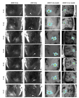 Axial T2-weighted MR images of the thalamotomy lesion at the AC-PC plane at 24 hours and 1 year posttreatment, and volumetric segmentation of the lesion at 24 hours posttreatment (represented on T1-weighted sequences). Axial (Ax) are views bounded by AC, PC, midline, and insular cortex. Orange indicates zone I (necrotic core); yellow, zone II (cytotoxic edema); and blue, zone III (vasogenic edema). All axial images are shown at the AC-PC plane. Horizontal line across coronal (Cor) images represents the AC-PC plane. Arrows indicate the residual lesion at 1 year. Pt = patient. |
Performance of Ultrafast DCE-MRI for Diagnosis of Prostate Cancer
|
Publication: Acad Radiol. 2018 Mar;25(3):349-58. PMID: 29167070 | PDF Authors: Chatterjee A, He D, Fan X, Wang S, Szasz T, Yousuf A, Pineda F, Antic T, Mathew M, Karczmar GS, Oto A. Institution: Department of Radiology, The University of Chicago, Chicago, IL, USA. Abstract: Rationale and Objectives: This study aimed to test high temporal resolution dynamic contrast-enhanced (DCE) magnetic resonance imaging (MRI) for different zones of the prostate and evaluate its performance in the diagnosis of prostate cancer (PCa). Determine whether the addition of ultrafast DCE-MRI improves the performance of multiparametric MRI. Materials and Methods: Patients (n = 20) with pathologically confirmed PCa underwent preoperative 3T MRI with T2-weighted, diffusion-weighted, and high temporal resolution (~2.2 seconds) DCE-MRI using gadoterate meglumine (Guerbet, Bloomington, IN) without an endorectal coil. DCE-MRI data were analyzed by fitting signal intensity with an empirical mathematical model to obtain parameters: percent signal enhancement, enhancement rate (α), washout rate (β), initial enhancement slope, and enhancement start time along with apparent diffusion coefficient (ADC) and T2 values. Regions of interests were placed on sites of prostatectomy verified malignancy (n = 46) and normal tissue (n = 71) from different zones. Results: Cancer (α = 6.45 ± 4.71 s-1, β = 0.067 ± 0.042 s-1, slope = 3.78 ± 1.90 s-1) showed significantly (P <.05) faster signal enhancement and washout rates than normal tissue (α = 3.0 ± 2.1 s-1, β = 0.034 ± 0.050 s-1, slope = 1.9 ± 1.4 s-1), but showed similar percentage signal enhancement and enhancement start time. Receiver operating characteristic analysis showed area under the curve for DCE parameters was comparable to ADC and T2 in the peripheral (DCE 0.67-0.82, ADC 0.80, T2 0.89) and transition zones (DCE 0.61-0.72, ADC 0.69, T2 0.75), but higher in the central zone (DCE 0.79-0.88, ADC 0.45, T2 0.45) and anterior fibromuscular stroma (DCE 0.86-0.89, ADC 0.35, T2 0.12). Importantly, combining DCE with ADC and T2 increased area under the curve by ~30%, further improving the diagnostic accuracy of PCa detection. Conclusion: Quantitative parameters from empirical mathematical model fits to ultrafast DCE-MRI improve diagnosis of PCa. DCE-MRI with higher temporal resolution may capture clinically useful information for PCa diagnosis that would be missed by low temporal resolution DCE-MRI. This new information could improve the performance of multiparametric MRI in PCa detection. "… MR images from different pulse sequences were registered using rigid registration in 3D Slicer." |
A Cost-Effective, In-House, Positioning and Cutting Guide System for Orthognathic Surgery
|
Publication: J Maxillofac Oral Surg. 2018 Mar;17(1):112-4. PMID: 29383005 | PDF Authors: McAllister P, Watson M, Burke E.
Abstract: INTRODUCTION: Technological advances in 3D printing can dramatically improve orthognathic surgical planning workflow. Custom positioning and cutting guides enable intraoperative reproduction of pre-planned osteotomy cuts and can result in greater surgical accuracy and patient safety. OBJECTIVES: This short paper describes the use of freeware (some with open-source) combined with in-house 3D printing facilities to produce reliable, affordable osteotomy cutting guides. METHODS: Open-source software, 3D Slicer, is used to visualise and segment three-dimensional planning models from imported conventional computed tomography (CT) scans. Freeware (Autodesk Meshmixer ©) allows digital manipulation of maxillary and mandibular components to plan precise osteotomy cuts. Bespoke cutting guides allow exact intraoperative positioning. These are printed in polylactic acid (PLA) using a fused-filament fabrication 3D printer. Fixation of the osteotomised segments is achieved using plating templates and four pre-adapted plates with planned screw holes over the thickest bone. We print maxilla/ mandible models with desired movements incorporated to use as a plating template. RESULTS: A 3D printer capable of reproducing a complete skull can be procured for £1000, with material costs in the region of £10 per case. Our production of models and guides typically takes less than 24 hours of total print time. The entire production process is frequently less than three days. Externally sourced models and guides cost significantly more, frequently encountering costs totalling £1500-£2000 for models and guides for a bimaxillary osteotomy. CONCLUSION: Three-dimensional guided surgical planning utilising custom cutting guides enables the surgeon to determine optimal orientation of osteotomy cuts and better predict the skeletal maxilla/mandible relationship following surgery. The learning curve to develop proficiency using planning software and printer settings is offset by increased surgical predictability and reduced theatre time, making this form of planning a worthy investment. |
Comparison of 3D Echocardiogram-Derived 3D Printed Valve Models to Molded Models for Simulated Repair of Pediatric Atrioventricular Valves
|
Publication: Pediatr Cardiol. 2018 Mar;39(3):538-47. PMID: 29181795 | PDF Authors: Scanlan AB, Nguyen AV, Ilina A, Lasso A, Cripe L, Jegatheeswaran A, Silvestro E, McGowan FX, Mascio CE, Fuller S, Spray TL, Cohen MS, Fichtinger G, Jolley MA. Institution: Department of Anesthesiology and Critical Care Medicine, Children's Hospital of Philadelphia, Philadelphia, PA, USA. Abstract: Mastering the technical skills required to perform pediatric cardiac valve surgery is challenging in part due to limited opportunity for practice. Transformation of 3D echocardiographic (echo) images of congenitally abnormal heart valves to realistic physical models could allow patient-specific simulation of surgical valve repair. We compared materials, processes, and costs for 3D printing and molding of patient-specific models for visualization and surgical simulation of congenitally abnormal heart valves. Pediatric atrioventricular valves (mitral, tricuspid, and common atrioventricular valve) were modeled from transthoracic 3D echo images using semi-automated methods implemented as custom modules in 3D Slicer. Valve models were then both 3D printed in soft materials and molded in silicone using 3D printed "negative" molds. using pre-defined assessment criteria, valve models were evaluated by congenital cardiac surgeons to determine suitability for simulation. Surgeon assessment indicated that the molded valves had superior material properties for the purposes of simulation compared to directly printed valves (p < 0.01). Patient-specific, 3D echo-derived molded valves are a step toward realistic simulation of complex valve repairs but require more time and labor to create than directly printed models. Patient-specific simulation of valve repair in children using such models may be useful for surgical training and simulation of complex congenital cases. |
A Preliminary Study on Precision Image Guidance for Electrode Placement in an EEG Study
|
Publication: Brain Topogr. 2018 Mar;31(2):174-85. PMID: 29204789 Authors: Trujillo P, Summers PE, Smith AK, Smith SA, Mainardi LT, Cerutti S, Claassen DO, Costa A. Institution: Department of Robotics Engineering, DGIST, Daegu, Republic of Korea. Abstract: Conventional methods for positioning electroencephalography electrodes according to the international 10/20 system are based on the manual identification of the principal 10/20 landmarks via visual inspection and palpation, inducing intersession variations in their determined locations due to structural ambiguity or poor visibility. To address the variation issue, we propose an image guidance system for precision electrode placement. Following the electrode placement according to the 10/20 system, affixed electrodes are laser-scanned together with the facial surface. For subsequent procedures, the laser scan is conducted likewise after positioning the electrodes in an arbitrary manner, and following the measurement of fiducial electrode locations, frame matching is performed to determine a transformation from the coordinate frame of the position tracker to that of the laser-scanned image. Finally, by registering the intra-procedural scan of the facial surface to the reference scan, the current tracking data of the electrodes can be visualized relative to the reference goal positions without manually measuring the four principal landmarks for each trial. The experimental results confirmed that use of the electrode navigation system significantly improved the electrode placement precision compared to the conventional 10/20 system (p < 0.005). The proposed system showed the possibility of precise image-guided electrode placement as an alternative to the conventional manual 10/20 system. "An open-source software package, 3D Slicer was used as a basic visualization platform" Funding:
|
Radiologic Factors Predicting Deterioration of Mental Status in Patients with Acute Traumatic Subdural Hematoma
|
Publication: World Neurosurg. 2018 Mar;111:e120-e134. PMID: 29248778 Authors: Won YD, Na MK, Ryu JI, Cheong JH, Kim JM, Kim CH, Han MH. Institution: Department of Neurosurgery, Hanyang University Guri Hospital, Gyonggi-do, Korea. Abstract: OBJECTIVE: To evaluate whether subdural hematoma (SDH) volume and other radiologic factors predict deterioration of mental status in patients with acute traumatic SDH. METHODS: SDH volumes were measured with a semiautomated tool. The area under the receiver operating characteristic curve was used to determine optimal cutoff values for mental deterioration, including the variables midline shift, SDH volume, hematoma thickness, and Sylvian fissure ratio. Multivariate logistic regression was used to calculate the odds ratio for mental deterioration based on several predictive factors. RESULTS: We enrolled 103 consecutive patients admitted to our hospital with acute traumatic SDH over an 8-year period. We observed an increase in SDH volume of approximately 7.2 mL as SDH thickness increased by 1 mm. A steeper slope for midline shift was observed in patients with SDH volumes of approximately 75 mL in the younger age group compared with patients in the older age group. When comparing cutoff values used to predict poor mental status at time of admission between the 2 age groups, we observed smaller midline shifts in the older patients. CONCLUSIONS: Among younger patients, an overall tendency for more rapid midline shift progression was observed in patients with relatively low SDH volumes compared with older patients. Older patients seem to tolerate larger hematoma volumes owing to brain atrophy compared with younger patients. When there is a midline shift, older patients seem to be more vulnerable to mental deterioration than younger patients. "We measured hematoma volume with 3D Slicer software." |
How to Precisely Measure the Volume Velocity Transfer Function of Physical Vocal Tract Models by External Excitation
|
Publication: PLoS One. 2018 Mar 15;13(3):e0193708. PMID: 29543829 | PDF Authors: Fleischer M, Mainka A, Kürbis S, Birkholz P. Institution: Division of Phoniatrics and Audiology, Department of Otorhinolaryngology, Faculty of Medicine Carl Gustav Carus, Technische Universität Dresden, Dresden, Germany. Abstract: Recently, 3D printing has been increasingly used to create physical models of the vocal tract with geometries obtained from magnetic resonance imaging. These printed models allow measuring the vocal tract transfer function, which is not reliably possible in vivo for the vocal tract of living humans. The transfer functions enable the detailed examination of the acoustic effects of specific articulatory strategies in speaking and singing, and the validation of acoustic plane-wave models for realistic vocal tract geometries in articulatory speech synthesis. To measure the acoustic transfer function of 3D-printed models, two techniques have been described: (1) excitation of the models with a broadband sound source at the glottis and measurement of the sound pressure radiated from the lips, and (2) excitation of the models with an external source in front of the lips and measurement of the sound pressure inside the models at the glottal end. The former method is more frequently used and more intuitive due to its similarity to speech production. However, the latter method avoids the intricate problem of constructing a suitable broadband glottal source and is therefore more effective. It has been shown to yield a transfer function similar, but not exactly equal to the volume velocity transfer function between the glottis and the lips, which is usually used to characterize vocal tract acoustics. Here, we revisit this method and show both, theoretically and experimentally, how it can be extended to yield the precise volume velocity transfer function of the vocal tract. |
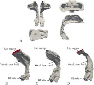 Printed and finite element models (A) Different views of the 3D-printed model of /a/. (B), (C) and (D) Finite element models of /a/, /u/ and /i/. For each model, the surface was partitioned into three regions representing the glottis, the lip region and the vocal tract walls. The merged voxel models for /a/, /u/ and /i/ were then converted back into triangle meshes using 3D Slicer. |
Longitudinal Microstructural Changes of Cerebral White Matter and their Association with Mobility Performance in Older Persons
|
Publication: PLoS One. 2018 Mar 19;13(3):e0194051. PMID: 29554115 | PDF Authors: Moscufo N, Wakefield DB, Meier DS, Cavallari M, Guttmann CRG, White WB3, Wolfson L. Institution: Center for Neurological Imaging, Department of Radiology, Brigham and Women's Hospital, Harvard Medical School, Boston, MA, USA. Abstract: Mobility impairment in older persons is associated with brain white matter hyperintensities (WMH), a common finding in magnetic resonance images and one established imaging biomarker of small vessel disease. The contribution of possible microstructural abnormalities within normal-appearing white matter (NAWM) to mobility, however, remains unclear. We used diffusion tensor imaging (DTI) measures, i.e. fractional anisotropy (FA), mean diffusivity (MD), axial diffusivity (AD), radial diffusivity (RD), to assess microstructural changes within supratentorial NAWM and WMH sub-compartments, and to investigate their association with changes in mobility performance, i.e. Tinetti assessment and the 2.5-meters walk time test. We analyzed baseline (N = 86, age ≥75 years) and 4-year (N = 41) follow-up data. Results from cross-sectional analysis on baseline data showed significant correlation between WMH volume and NAWM-FA (r = -0.33, p = 0.002), NAWM-AD (r = 0.32, p = 0.003) and NAWM-RD (r = 0.39, p = 0.0002). Our longitudinal analysis showed that after 4-years, FA and AD decreased and RD increased within NAWM. In regional tract-based analysis decrease in NAWM-FA and increase in NAWM-RD within the genu of the corpus callosum correlated with slower walk time independent of age, gender and WMH burden. In conclusion, global DTI indices of microstructural integrity indicate that significant changes occur in the supratentorial NAWM over four years. The observed changes likely reflect white matter deterioration resulting from aging as well as accrual of cerebrovascular injury associated with small vessel disease. The observed association between mobility scores and regional measures of NAWM microstructural integrity within the corpus callosum suggests that subtle changes within this structure may contribute to mobility impairment. Funding:
|
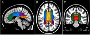 Relative spatial location of the regions of interest analyzed. Three-dimensional models of the sub-regions of the corpus callosum and corona radiata are illustrated. Models were produced in 3D Slicer using the MNI white matter parcellation map and they are shown overlaid on the MNI brain in sagittal (A), axial (B) and coronal (C) views. Regions shown are the anterior corona radiata (ACR, green), superior corona radiata (SCR, blue), posterior corona radiata (PCR, purple), genu of corpus callosum (GCC, red), body of corpus callosum (BCC, yellow), and splenium of corpus callosum (SCC, orange). Views are from left (A), top (B) and front (C) of head. For the regional assessment of the diffusion indices we used a mask with skeletonized ROIs in the subject native space. |
Micro-computed Tomographic Evaluation of the Shaping Ability of XP-endo Shaper, iRaCe, and EdgeFile Systems in Long Oval-shaped Canals
|
Publication: J Endod. 2018 Mar;44(3):489-95. PMID: 29273492 Authors: Versiani MA, Carvalho KKT, Mazzi-Chaves JF, Sousa-Neto MD. Institution: Department of Restorative Dentistry, Dental School of Ribeirão Preto, University of São Paulo, Brazil. Abstract: INTRODUCTION: This study evaluated the shaping ability of the XP-endo Shaper (FKG Dentaire SA, La Chaux-de-Fonds, Switzerland), iRaCe (FKG Dentaire SA), and EdgeFile (EdgeEndo, Albuquerque, NM) systems using micro-computed tomographic (micro-CT) technology. METHODS: Thirty long oval-shaped canals from mandibular incisors were matched anatomically using micro-CT scanning (SkyScan1174v2; Bruker-microCT, Kontich, Belgium) and distributed into 3 groups (n = 10) according to the canal preparation protocol (ie, XP-endo Shaper, iRaCe, and EdgeFile systems). Coregistered images, before and after preparation, were evaluated for morphometric measurements of the volume, surface area, structure model index (SMI), untouched walls, area, perimeter, roundness, and diameter. Data were statistically compared between groups using the 1-way analysis of variance post hoc Tukey test and within groups with the paired sample t test (α = 5%). RESULTS: Within groups, preparation significantly increased all tested parameters (P < .05). No statistical difference was observed in the mean percentage increase of the volume (〜52%) and surface area (10.8%-14.2%) or the mean percentage of the remaining unprepared canal walls between groups (8.17%-9.83%) (P > .05). The XP-endo Shaper significantly altered the overall geometry of the root canal to a more conical shape (SMI = 2.59) when compared with the other groups (P < .05). After preparation protocols, changes in area, perimeter, roundness, and minor and major diameters of the root canals in the 5 mm of the root apex showed no difference between groups (P > .05). CONCLUSIONS: The XP-endo Shaper, iRaCe, and EdgeFile systems showed a similar shaping ability. Despite the XP-endo Shaper had significantly altered the overall geometry of the root canal to a more conical shape, neither technique was capable of completely preparing the long oval-shaped canals of mandibular incisors. "...models of the canals were coregistered with their respective preoperative data sets using the rigid registration module of the 3D Slicer v.4.3.1 software" |
Cerebral Radiation Necrosis: An Analysis of Clinical and Quantitative Imaging and Volumetric Features
|
Publication: World Neurosurg. 2018 Mar;111:e485-e494. PMID: 29288110 Authors: Feng R, Loewenstern J, Aggarwal A, Pawha P, Gilani A, Iloreta AM, Bakst R, Miles B, Bederson J, Costa A, Gupta V, Shrivastava R. Abstract: BACKGROUND: Radiation therapy (RT) is an effective treatment for primary brain tumors and intracranial metastases, but can occasionally precede new enhancing lesions on imaging studies that are difficult to discern between a tumor recurrence (TR) or radiation necrosis (RN). There is a need to identify clinical presentation and imaging patterns that may obviate the need for invasive definitive biopsy. OBJECTIVE: To describe clinical and imaging characteristics of RN lesions compared to those from a TR. METHODS: Patients who received RT and subsequently presented with a new intracranial lesion were reviewed from 2001-2016. Twenty-seven patients were identified with adequate records and confirmed pathology to have RN present or TR only. Patient and lesion characteristics were assessed utilizing univariate and multivariate logistic regression analyses. Sensitivity and specificities were calculated for imaging features and quantitatively segmented lesion and edema volumes for identifying RN. RESULTS: Karnofsky Performance Status (KPS) at presentation significantly predicted pathological diagnosis on univariate analysis (p = 0.044). Radiation dosage and time from RT to lesion onset did not differ among pathological diagnosis groups. No differences existed between RN and TR on quantitative imaging analyses. Multivariate logistic regression found higher KPS to be an independent factor associated with TR relative to RN (OR = 1.26, 95% CI = 1.02-1.56, p = 0.030). CONCLUSIONS: Diagnostic imaging can often be inaccurate in detecting RN alone, even with quantitative volume assessment. Functional status on re-presentation may increase the likelihood of accurate diagnosis prior to a definitive biopsy when neuroimaging remains unclear. "...segmented for contrast-enhancement, lesion, and edema volumes using a semi-automatic image processing 3D Slicer software v.4.6." |
Nonlinear Deformation of Tractography in Ultrasound-guided Low-grade Gliomas Resection
|
Publication: Int J Comput Assist Radiol Surg. 2018 Mar;13(3):457-67. PMID: 29299739 Authors: Xiao Y, Eikenes L, Reinertsen I, Rivaz H. Institution: PERFORM Centre, Concordia University, Montreal, Canada. Abstract: Purpose: In brain tumor surgeries, maximum removal of cancerous tissue without compromising normal brain functions can improve the patient's survival rate and therapeutic benefits. To achieve this, diffusion MRI and intra-operative ultrasound (iUS) can be highly instrumental. While diffusion MRI allows the visualization of white matter tracts and helps define the resection plan to best preserve the eloquent areas, iUS can effectively track the brain shift after craniotomy that often renders the pre-surgical plan invalid, ensuring the accuracy and safety of the intervention. Unfortunately, brain shift correction using iUS and automatic registration has never been shown for brain tractography so far despite its rising significance in brain tumor resection. Methods: We employed a correlation-ratio-based nonlinear registration algorithm to account for brain shift through MRI-iUS registration and used the recovered deformations to warp both the brain anatomy and tractography seen in pre-surgical plans. The overall technique was demonstrated retrospectively on four patients who underwent iUS-guided low-grade brain gliomas resection. Results: Through qualitative and quantitative evaluations, the preoperative MRI and iUS scans were well realigned after nonlinear registration, and the deformed brain tumor volumes and white matter tracts showed large displacements away from the pre-surgical plans. Conclusions: We are the first to demonstrate the technique to track nonlinear deformation of brain tractography using real clinical MRI and iUS data, and the results confirm the need for updating white matter tracts due to tissue shift during surgery. "The results were visualized with the 3D Slicer software" |
Lateral Ventricular Volume Asymmetry Predicts Poor Outcome After Spontaneous Intracerebral Hemorrhage
|
Publication: World Neurosurg. 2018 Feb;110:e958-e964. PMID: 29203311 Authors: Chen J, Zhang D, Li Z, Dong Y, Han K, Wang J, Hou L. Institution: Department of Neurosurgery, Shanghai Institute of Neurosurgery, PLA Institute of Neurosurgery, Changzheng Hospital, Second Military Medical University, Shanghai, China. Abstract: Background: Midline shift (MLS) has been a known predictor for prognosis after spontaneous intracerebral hemorrhage (ICH), whereas it is secondary to lateral ventricular compression. In this study, we investigated whether lateral ventricular volume (LVV) asymmetry caused by ventricular compression was independently associated with poor outcome of ICH. Methods: We retrospectively studied clinical patients with spontaneous ICH from January 2010 to January 2017. LVV was calculated using slicer software. LVV ratio (LVR) was then determined to quantitatively evaluate LVV asymmetry, and its relationship with poor outcome was tested by logistic regression model. Receiver operating characteristic (ROC) curve analysis was performed to identify the optimized baseline LVR cutoff point to predict poor outcome. Results: 188 patients were included, of whom 41% (77/188) experienced a poor outcome. Multivariate logistic regression analysis identified baseline LVR as an independent predictor for poor outcome after ICH. The predictive value of baseline LVR was confirmed by ROC analysis (area under the curve = 0.742; P < 0.001). The optimized baseline LVR cutoff point was 3.7, with a sensitivity of 64.9% and specificity of 80.2%. using LVR >3.7 as an exposure factor yielded an odds ratio of 7.49 (P < 0.001), and a risk ratio of 2.98 (P < 0.001). Conclusions: LVV asymmetry was associated with clinical prognosis after ICH, and high LVR (>3.7) might independently predict poor outcome. "LVV and HV measurements were conducted using three dimensional 3D Slicer v. 4.0." |
High Fidelity Virtual Reality Orthognathic Surgery Simulator
|
Publication: Proc SPIE Int Soc Opt Eng. 2018 Feb;10576. PMID: 29977103 | PDF Authors: Arikatla VS, Tyagi M, Enquobahrie A, Nguyen T, Blakey GH, White R, Paniagua B. Institution: Kitware Inc., Carrboro, NC, USA. Abstract: Surgical simulators are powerful tools that assist in providing advanced training for complex craniofacial surgical procedures and objective skills assessment such as the ones needed to perform Bilateral Sagittal Split Osteotomy (BSSO). One of the crucial steps in simulating BSSO is accurately cutting the mandible in a specific area of the jaw, where surgeons rely on high fidelity visual and haptic cues. In this paper, we present methods to simulate drilling and cutting of the bone using the burr and the motorized oscillating saw respectively. Our method allows low computational cost bone drilling or cutting while providing high fidelity haptic feedback that is suitable for real-time virtual surgery simulation. Funding:
|
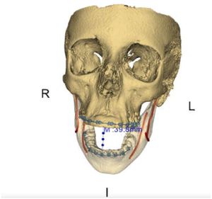 High-resolution model (obtained from CBCT) that illustrates the pre-surgical state of BSSO, with mandible rendered in full opening width. The model includes 3-dimensional reconstruction of craniofacial hard tissue (bone, in solid/semitransparent yellow) and soft tissue (vessels in solid red, gingiva in semitransparent pink) structures, rendered altogether with orthodontic appliances (brackets in solid blue). "In order to generate the 3D model of the bony structures of the patient we employed 3D Slicer Segmentation Module to threshold the scan of the patient." |
Volumetric Analysis of Magnetic Resonance-guided Focused Ultrasound Thalamotomy Lesions
|
Publication: Neurosurg Focus. 2018 Feb;44(2):E6. PMID: 29385921 | PDF Authors: Harary M, Essayed WI, Valdes PA, McDannold N, Cosgrove GR. Institution: Departments of Neurosurgery and Radiology, Brigham and Women's Hospital, Harvard Medical School, Boston, MA, USA. Abstract: OBJECTIVE Magnetic resonance-guided focused ultrasound (MRgFUS) thalamotomy was recently approved for use in the treatment of medication-refractory essential tremor (ET). Previous work has described lesion appearance and volume on MRI up to 6 months after treatment. Here, the authors report on the volumetric segmentation of the thalamotomy lesion and associated edema in the immediate postoperative period and 1 year following treatment, and relate these radiographic characteristics with clinical outcome. METHODS Seven patients with medication-refractory ET underwent MRgFUS thalamotomy at Brigham and Women's Hospital and were monitored clinically for 1 year posttreatment. Treatment effect was measured using the Clinical Rating Scale for Tremor (CRST). MRI was performed immediately postoperatively, 24 hours posttreatment, and at 1 year. Lesion location and the volumes of the necrotic core (zone I) and surrounding edema (cytotoxic, zone II; vasogenic, zone III) were measured on thin-slice T2-weighted images using 3D Slicer software. RESULTS Patients had significant improvement in overall CRST scores (baseline 51.4 ± 10.8 to 24.9 ± 11.0 at 1 year, p = 0.001). The most common adverse events (AEs) in the 1-month posttreatment period were transient gait disturbance (6 patients) and paresthesia (3 patients). The center of zone I immediately posttreatment was 5.61 ± 0.9 mm anterior to the posterior commissure, 14.6 ± 0.8 mm lateral to midline, and 11.0 ± 0.5 mm lateral to the border of the third ventricle on the anterior commissure-posterior commissure plane. Zone I, II, and III volumes immediately posttreatment were 0.01 ± 0.01, 0.05 ± 0.02, and 0.33 ± 0.21 cm3, respectively. These volumes increased significantly over the first 24 hours following surgery. The edema did not spread evenly, with more notable expansion in the superoinferior and lateral directions. The spread of edema inferiorly was associated with the incidence of gait disturbance. At 1 year, the remaining lesion location and size were comparable to those of zone I immediately posttreatment. Zone volumes were not associated with clinical efficacy in a statistically significant way. CONCLUSIONS MRgFUS thalamotomy demonstrates sustained clinical efficacy at 1 year for the treatment of medication-refractory ET. This technology can create accurate, predictable, and small-volume lesions that are stable over time. Instances of AEs are transient and are associated with the pattern of perilesional edema expansion. Additional analysis of a larger MRgFUS thalamotomy cohort could provide more information to maximize clinical effect and reduce the rate of long-lasting AEs. |
 Axial T2-weighted MR images of the thalamotomy lesion at the AC-PC plane at 24 hours and 1 year posttreatment, and volumetric segmentation of the lesion at 24 hours posttreatment (represented on T1-weighted sequences). Axial (Ax) are views bounded by AC, PC, midline, and insular cortex. Orange indicates zone I (necrotic core); yellow, zone II (cytotoxic edema); and blue, zone III (vasogenic edema). All axial images are shown at the AC-PC plane. Horizontal line across coronal (Cor) images represents the AC-PC plane. Arrows indicate the residual lesion at 1 year. Pt = patient. |
SVA: Shape Variation Analyzer
|
Publication: Proc SPIE Int Soc Opt Eng. 2018 Feb;10578. PMID: 29780198 | PDF Authors: de Dumast P, Mirabel C, Paniagua B, Yatabe M, Ruellas A, Tubau N, Styner M, Cevidanes L, Prieto JC. Institution: University of Michigan, Ann Arbor, MI, USA. Abstract: Temporo-mandibular osteo arthritis (TMJ OA) is characterized by progressive cartilage degradation and subchondral bone remodeling. The causes of this pathology remain unclear. Current research efforts are concentrated in finding new biomarkers that will help us understand disease progression and ultimately improve the treatment of the disease. In this work, we present Shape Variation Analyzer (SVA), the goal is to develop a noninvasive technique to provide information about shape changes in TMJ OA. SVA uses neural networks to classify morphological variations of 3D models of the mandibular condyle. The shape features used for training include normal vectors, curvature and distances to average models of the condyles. The selected features are purely geometric and are shown to favor the classification task into 6 groups generated by consensus between two clinician experts. With this new approach, we were able to accurately classify 3D models of condyles. In this paper, we present the methods used and the results obtained with this new tool. Funding:
|
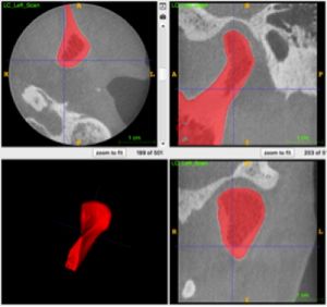 Manual segmentation of Condyles using ITK-SNAP. "Using EasyClip, a module included in 3D Slicer, the condylar models are simultaneously cropped to define the condylar region of interest. The cropped segmentation maps are used to generated regular tessellations." |
Automatic Quantification Framework to Detect Cracks in Teeth
|
Publication: Proc SPIE Int Soc Opt Eng. 2018 Feb;10578. PMID: 29769755 | PDF Authors: Shah H, Hernandez P, Budin F, Chittajallu D, Vimort JB, Walters R, Mol A, Khan A, Paniagua B. Institution: Kitware, Inc., Carrboro, NC, USA. Abstract: Studies show that cracked teeth are the third most common cause for tooth loss in industrialized countries. If detected early and accurately, patients can retain their teeth for a longer time. Most cracks are not detected early because of the discontinuous symptoms and lack of good diagnostic tools. Currently used imaging modalities like Cone Beam Computed Tomography (CBCT) and intraoral radiography often have low sensitivity and do not show cracks clearly. This paper introduces a novel method that can detect, quantify, and localize cracks automatically in high resolution CBCT (hr-CBCT) scans of teeth using steerable wavelets and learning methods. These initial results were created using hr-CBCT scans of a set of healthy teeth and of teeth with simulated longitudinal cracks. The cracks were simulated using multiple orientations. The crack detection was trained on the most significant wavelet coefficients at each scale using a bagged classifier of Support Vector Machines. Our results show high discriminative specificity and sensitivity of this method. The framework aims to be automatic, reproducible, and open-source. Future work will focus on the clinical validation of the proposed techniques on different types of cracks ex-vivo. We believe that this work will ultimately lead to improved tracking and detection of cracks allowing for longer lasting healthy teeth. Funding:
|
 Example of synthetic simulated dataset for a (a) healthy second molar, and(c) simulated crack sample. Data was post-processed to improve realism. (b) Healthy second molar after smoothing and (d) simulated crack sample after smoothing. Arrow points to the simulated crack. "Relevant anatomy of the teeth was segmented using 3D Slicer Segment Editor." |
Detection of Bone Loss via Subchondral Bone Analysis
|
Publication: Proc SPIE Int Soc Opt Eng. 2018 Feb;10578. PMID: 29769754 | PDF Authors: Vimort JB, Ruellas A, Prothero J, Marron JS, McCormick M, Cevidanes L, Benavides E, Paniagua B. Institution: Kitware, Inc., Carrboro, NC, USA. Abstract: To date, there is no single sign, symptom, or test that can clearly diagnose early stages of Temporomandibular Joint Osteoarthritis (TMJ OA). However, it has been observed that changes in the bone occur in early stages of this disease, involving structural changes both in the texture and morphometry of the bone marrow and the subchondral cortical plate. In this paper we present a tool to detect and highlight subtle variations in subchondral bone structure obtained from high resolution Cone Beam Computed Tomography (hr-CBCT) in order to help with detecting early TMJ OA. The proposed tool was developed in ITK and 3D Slicer and it has been disseminated as open-source software tools. We have validated both our texture analysis and morphometry analysis biomarkers for detection of TMJ OA comparing hr-CBCT to μCT. Our initial statistical results using the multidimensional features computed with our tool indicate that it is possible to classify areas of demonstrated loss of trabecular bone in both μCT and hr-CBCT. This paper describes the first steps to alleviate the current inability of radiological changes to diagnose TMJ OA before morphological changes are too advanced by quantifying subchondral bone biomarkers. This paper indicates that texture based and morphometry based biomarkers have the potential to identify OA patients at risk for further bone destruction. Funding:
|
Development of a Patient-specific Tumor Mold using Magnetic Resonance Imaging and 3-Dimensional Printing Technology for Targeted Tissue Procurement and Radiomics Analysis of Renal Masses
|
Publication: Urology. 2018 Feb;112:209-14. PMID: 29056576 | PDF Authors: Dwivedi DK, Chatzinoff Y, Zhang Y, Yuan Q, Fulkerson M, Chopra R, Brugarolas J, Cadeddu JA, Kapur P, Pedrosa I. Institution: Department of Radiology, UT Southwestern Medical Center, Dallas, TX, USA. Abstract: Objective: To implement a platform for colocalization of in vivo quantitative multiparametric magnetic resonance imaging features with ex vivo surgical specimens of patients with renal masses using patient-specific 3-dimensional (3D)-printed tumor molds, which may aid in targeted tissue procurement and radiomics and radiogenomic analyses. Materials and Methods: Volumetric segmentation of 6 renal masses was performed with 3D Slicer to create a 3D tumor model. A slicing guide template was created with specialized software, which included notches corresponding to the anatomic locations of the magnetic resonance images. The tumor model was subtracted from the slicing guide to create a depression in the slicing guide corresponding to the exact size and shape of the tumor. A customized, tumor-specific, slicing guide was then printed using a 3D printer. After partial nephrectomy, the surgical specimen was bivalved through the preselected magnetic resonance imaging (MRI) plane. A thick slab of the tumor was obtained, fixed, and processed as a whole-mount slide and was correlated to multiparametric MRI findings. Results: All patients successfully underwent partial nephrectomy and adequate fitting of the tumor specimens within the 3D mold was achieved in all tumors. Distinct in vivo MRI features corresponded to unique pathologic characteristics in the same tumor. The average cost of printing each mold was US$160.7 ± 111.1 (range: US$20.9-$350.7). Conclusion: MRI-based preoperative 3D printing of tumor-specific molds allow for accurate sectioning of the tumor after surgical resection and colocalization of in vivo imaging features with tissue-based analysis in radiomics and radiogenomic studies. |
A Surface-based Approach to Determine Key Spatial Parameters of the Acetabulum in a Standardized Pelvic Coordinate System
|
Publication: Med Eng Phys. 2018 Feb;52:22-30. PMID: 29269225 | PDF Authors: Chen X, Jia P, Wang Y, Zhang H, Wang L, Frangi AF, Taylor ZA. Institution: Institute of Biomedical Manufacturing and Life Quality Engineering, School of Mechanical Engineering, Shanghai Jiaotong University, Shanghai, China. Abstract: Accurately determining the spatial relationship between the pelvis and acetabulum is challenging due to their inherently complex three-dimensional (3D) anatomy. A standardized 3D pelvic coordinate system (PCS) and the precise assessment of acetabular orientation would enable the relationship to be determined. We present a surface-based method to establish a reliable PCS and develop software for semi-automatic measurement of acetabular spatial parameters. Vertices on the acetabular rim were manually extracted as an eigenpoint set after 3D models were imported into the software. A reliable PCS consisting of the anterior pelvic plane, midsagittal pelvic plane, and transverse pelvic plane was then computed by iteration on mesh data. A spatial circle was fitted as a succinct description of the acetabular rim. Finally, a series of mutual spatial parameters between the pelvis and acetabulum were determined semi-automatically, including the center of rotation, radius, and acetabular orientation. Pelvic models were reconstructed based on high-resolution computed tomography images. Inter- and intra-rater correlations for measurements of mutual spatial parameters were almost perfect, showing our method affords very reproducible measurements. The approach will thus be useful for analyzing anatomic data and has potential applications for preoperative planning in individuals receiving total hip arthroplasty. "Surface models are reconstructed from computed tomography (CT) data volumes through the threshold and region-growing segmentation method using 3D Slicer v.4.2." |
Imaging of Concussion in Young Athletes
|
Publication: Neuroimaging Clin N Am. 2018 Feb;28(1):43-53. PMID: 29157852 | PDF Authors: Guenette JP, Shenton ME, Koerte IK. Institution: Department of Radiology, Brigham and Women's Hospital, Harvard Medical School, Boston, MA, USA. Abstract: Conventional neuroimaging examinations are typically normal in concussed young athletes. A current focus of research is the characterization of subtle abnormalities after concussion using advanced neuroimaging techniques. These techniques have the potential to identify biomarkers of concussion. In the future, such biomarkers will likely provide important clinical information regarding the appropriate time interval before return to play, as well as the risk for prolonged postconcussive symptoms and long-term cognitive impairment. This article discusses results from advanced imaging techniques and emphasizes imaging modalities that will likely become available in the near future for the clinical evaluation of concussed young athletes. "using a tool such as 3D Slicer (Surgical Planning Laboratory, Brigham and Women's Hospital, Boston, MA), regions of interest within the brain can be manually segmented or parcellated."
|
Advantages and Disadvantages in Image Processing with Free Software in Radiology
|
Publication: J Med Syst. 2018 Jan 15;42(3):36. PMID: 29333590 Authors: Mujika KM, Méndez JAJ, de Miguel AF.
Abstract: Currently, there are sophisticated applications that make it possible to visualize medical images and even to manipulate them. These software applications are of great interest, both from a teaching and a radiological perspective. In addition, some of these applications are known as Free Open Source Software because they are free and the source code is freely available, and therefore it can be easily obtained even on personal computers. Two examples of free open source software are Osirix Lite® and 3D Slicer®. However, this last group of free applications have limitations in its use. For the radiological field, manipulating and post-processing images is increasingly important. Consequently, sophisticated computing tools that combine software and hardware to process medical images are needed. In radiology, graphic workstations allow their users to process, review, analyse, communicate and exchange multidimensional digital images acquired with different image-capturing radiological devices. These radiological devices are basically CT (Computerised Tomography), MRI (Magnetic Resonance Imaging), PET (Positron Emission Tomography), etc. Nevertheless, the programs included in these workstations have a high cost which always depends on the software provider and is always subject to its norms and requirements. With this study, we aim to present the advantages and disadvantages of these radiological image visualization systems in the advanced management of radiological studies. We will compare the features of the VITREA2® and AW VolumeShare 5® radiology workstation with free open source software applications like OsiriX® and 3D Slicer®, with examples from specific studies. |
WRIST: A WRist Image Segmentation Toolkit for Carpal bone Delineation from MRI
|
Publication: Comput Med Imaging Graph. 2018 Jan;63:31-40. PMID: 29331208 | PDF Authors: Foster B, Joshi AA, Borgese M, Abdelhafez Y, Boutin RD, Chaudhari A. Institution: Department of Biomedical Engineering, University of California Davis, Davis, CA, USA. Abstract: Segmentation of the carpal bones from 3D imaging modalities, such as magnetic resonance imaging (MRI), is commonly performed for in vivo analysis of wrist morphology, kinematics, and biomechanics. This crucial task is typically carried out manually and is labor intensive, time consuming, subject to high inter- and intra-observer variability, and may result in topologically incorrect surfaces. We present a method, WRist Image Segmentation Toolkit (WRIST), for 3D semi-automated, rapid segmentation of the carpal bones of the wrist from MRI. In our method, the boundary of the bones were iteratively found using prior known anatomical constraints and a shape-detection level set. The parameters of the method were optimized using a training dataset of 48 manually segmented carpal bones and evaluated on 112 carpal bones which included both healthy participants without known wrist conditions and participants with thumb basilar osteoarthritis (OA). Manual segmentation by two expert human observers was considered as a reference. On the healthy subject dataset we obtained a Dice overlap of 93.0 ± 3.8, Jaccard Index of 87.3 ± 6.2, and a Hausdorff distance of 2.7 ± 3.4 mm, while on the OA dataset we obtained a Dice overlap of 90.7 ± 8.6, Jaccard Index of 83.0 ± 10.6, and a Hausdorff distance of 4.0 ± 4.4 mm. The short computational time of 20.8 s per bone (or 5.1 s per bone in the parallelized version) and the high agreement with the expert observers gives WRIST the potential to be utilized in musculoskeletal research. v.4.6." "Our work aimed at providing improved segmentation accuracy compared to existing methods while also having significantly decreased computational time. It was implemented as an open-source module for the popular image analysis tool, 3D Slicer." |
Enhanced Preoperative Deep Inferior Epigastric Artery Perforator Flap Planning with a 3D-Printed Perforasome Template: Technique and Case Report
|
Publication: Brachytherapy. Plast Reconstr Surg Glob Open. 2018 Jan 23;6(1):e1644. PMID: 29464169 | PDF Authors: Chae MP, Hunter-Smith DJ, Rostek M, Smith JA, Rozen WM. Institution: Department of Surgery, School of Clinical Sciences at Monash Health, Monash University, Monash Medical Centre, Clayton, Victoria, Australia. Abstract: Optimizing preoperative planning is widely sought in deep inferior epigastric artery perforator (DIEP) flap surgery. One reason for this is that rates of fat necrosis remain relatively high (up to 35%), and that adjusting flap design by an improved understanding of individual perforasomes and perfusion characteristics may be useful in reducing the risk of fat necrosis. Imaging techniques have substantially improved over the past decade, and with recent advances in 3D printing, an improved demonstration of imaged anatomy has become available. We describe a 3D-printed template that can be used preoperatively to mark out a patient's individualized perforasome for flap planning in DIEP flap surgery. We describe this "perforasome template" technique in a case of a 46-year-old woman undergoing immediate unilateral breast reconstruction with a DIEP flap. Routine preoperative computed tomographic angiography was performed, with open-source software (3D Slicer, Autodesk MeshMixer and Cura) and a desktop 3D printer (Ultimaker 3E) used to create a template used to mark intra-flap, subcutaneous branches of deep inferior epigastric artery (DIEA) perforators on the abdomen. An individualized 3D printed template was used to estimate the size and boundaries of a perforasome and perfusion map. The information was used to aid flap design. We describe a new technique of 3D printing a patient-specific perforasome template that can be used preoperatively to infer perforasomes and aid flap design. |
DCE-MRI Pharmacokinetic-Based Phenotyping of Invasive Ductal Carcinoma: A Radiomic Study for Prediction of Histological Outcomes
|
Publication: Contrast Media Mol Imaging. 2018 Jan 17;2018:5076269. PMID: 29581709 | PDF Authors: Monti S, Aiello M, Incoronato M, Grimaldi AM, Moscarino M, Mirabelli P, Ferbo U, Cavaliere C, Salvatore M. Institution: IRCCS SDN, Naples, Italy. Abstract: Breast cancer is a disease affecting an increasing number of women worldwide. Several efforts have been made in the last years to identify imaging biomarker and to develop noninvasive diagnostic tools for breast tumor characterization and monitoring, which could help in patients' stratification, outcome prediction, and treatment personalization. In particular, radiomic approaches have paved the way to the study of the cancer imaging phenotypes. In this work, a group of 49 patients with diagnosis of invasive ductal carcinoma was studied. The purpose of this study was to select radiomic features extracted from a DCE-MRI pharmacokinetic protocol, including quantitative maps of ktrans, kep, ve, iAUC, and R1 and to construct predictive models for the discrimination of molecular receptor status (ER+/ER-, PR+/PR-, and HER2+/HER2-), triple negative (TN)/non-triple negative (NTN), ki67 levels, and tumor grade. A total of 163 features were obtained and, after feature set reduction step, followed by feature selection and prediction performance estimations, the predictive model coefficients were computed for each classification task. The AUC values obtained were 0.826 ± 0.006 for ER+/ER-, 0.875 ± 0.009 for PR+/PR-, 0.838 ± 0.006 for HER2+/HER2-, 0.876 ± 0.007 for TN/NTN, 0.811 ± 0.005 for ki67+/ki67-, and 0.895 ± 0.006 for lowGrade/highGrade. In conclusion, DCE-MRI pharmacokinetic-based phenotyping shows promising for discrimination of the histological outcomes. "... dynamic motion-corrected sequence and the bounding box were given as input to the SegmentCAD module of dynamic motion-corrected sequence and the bounding box were given as input to the SegmentCAD module of 3D Slicer, which automatically segmented the lesion on the basis of the temporal dynamic of the signal." |
MRI-Based Experimentations of Fingertip Flat Compression: Geometrical Measurements and Finite Element Inverse Simulations to Investigate Material Property Parameters
|
Publication: J Biomech. 2018 Jan 23;67:166-71. PMID: 29217092 Authors: Dallard J, Merlhiot X, Petitjean N, Duprey S. Institution: University of Lyon, France. Abstract: Modeling human-object interactions is a necessary step in the ergonomic assessment of products. Fingertip finite element models can help investigating these interactions, if they are built based on realistic geometrical data and material properties. The aim of this study was to investigate the fingertip geometry and its mechanical response under compression, and to identify the parameters of a hyperelastic material property associated to the fingertip soft tissues. Fingertip compression tests in an MRI device were performed on 5 subjects at either 2 or 4 N and at 15° or 50°. The MRI images allowed to document both the internal and external fingertip dimensions and to build 5 subject-specific finite element models. Simulations reproducing the fingertip compression tests were run to obtain the material property parameters of the soft tissues. results indicated that two ellipses in the sagittal and longitudinal plane could describe the external fingertip geometry. The internal geometries indicated an averaged maximal thickness of soft tissues of 6.4 ± 0.8 mm and a 4 ± 1 mm height for the phalanx bone. The averaged deflections under loading went from 1.8 ± 0.3 mm at 2 N, 50° to 3.1 ± 0.2 mm at 4 N, 15°. Finally, the following set of parameters for a second order hyperelastic law to model the fingertip soft tissues was proposed: C01=0.59 ± 0.09 kPa and C20 = 2.65 ± 0.88 kPa. These data should facilitate further efforts on fingertip finite element modeling. "For each MRI acquisition, manual segmentations were performed using 3D Slicer. |
Objective Assessment of Colonoscope Manipulation Skills in Colonoscopy Training
|
Publication: Int J Comput Assist Radiol Surg. 2018 Jan;13(1):105-14. PMID: 29086234 Authors: Holden MS, Wang CN, MacNeil K, Church B, Hookey L, Fichtinger G, Ungi T. Institution: Laboratory for Percutaneous Surgery, School of Computing, Queen's University, Kingston, ON, Canada. Abstract: Objective: Manipulation of the colonoscope is a technical challenge for novice clinicians which is best learned in a simulated environment. It involves the coordination of scope tip steering with scope insertion, using a rotated image as reference. The purpose of this work is to develop and validate a system which objectively assesses colonoscopy technical skills proficiency in an arbitrary training environment, allowing novices to assess their technical proficiency prior to real patient encounters. Methods: We implemented a motion tracking setup to objectively analyze and assess the way operators perform colonoscopies, including an analysis of wrist and elbow joint motions. Subsequently, we conducted a validation study to verify whether our motion analysis could discriminate novice colonoscopists from experts. Participants navigated a wooden bench-top model using a standard colonoscope while their motions were tracked. Results: The developed motion tracking setup allowed colonoscopists of varying levels of proficiency to have their colonoscope manipulation assessed, and was able to be operated by a trained non-technical operator. Novice operators had significantly greater median times (101.5 vs. 31.5 s) and number of hand movements (62.0 vs. 21.5) than experts. Experts, however, spent a significantly greater proportion of time in extreme ranges of wrist and elbow joint motion than novices. Conclusion: We have developed and implemented a hand and joint motion analysis system that is able to discriminate novices from experts based on objective measures of motion. These metrics could, thus, serve as proxies for technical proficiency during training. "The PLUS software library was used to acquire and send the tracking data to the 3D Slicer software." |
Primary Trigeminal Neuralgia is Associated with Posterior Fossa Crowdedness: A Prospective Case-Control Study
|
Publication: J Clin Neurosci. 2018 Jan;47:89-92. PMID: 29066228 Authors: Cheng J, Meng J, Liu W, Zhang H, Hui X, Lei D. Institution: Department of Neurosurgery, West China Hospital, Sichuan University, Chengdu, China. Abstract: Neurovascular conflict (NVC) has been postulated to be the underlying cause of trigeminal neuralgia (TN). Does the posterior fossa crowdedness increase the chance of NVC? The aim of this study was to quantitatively measure the posterior fossa crowdedness in patients with TN and to perform a comparison with healthy controls. We conducted a prospective case-control study of 46 patients diagnosed with primary TN and 46 sex- and age-matched healthy controls. All subjects underwent high-resolution three-dimensional MRI, and the 3D Slicer software was used to measure the posterior fossa volume (PFV) and hindbrain volume (HBV). The posterior fossa crowdedness index (PFCI) was calculated as HBV/PFV × 100%. The results showed that patients with TN had a larger HBV (155.4 ± 23.2 cm3 versus 152.9 ± 13.5 cm3, P = .16) and a smaller PFV (182.7 ± 18.3 cm3 versus 186.1 ± 11.7 cm3, P = .42) as compared to control subjects, but these values were not significantly different. The mean PFCI was significantly higher in patients with TN than in controls (85.1% ± 3.4% versus 82.2% ± 5.3%; P = .03). Women had a more crowded posterior fossa than men (85.8% ± 2.1% versus 84.1% ± 2.6%; P = .023) in patients with TN. The correlation analysis showed that a higher PFCI was associated with younger age (P = .02), woman (P = .014), and TN disease (P = .001). From this study, we conclude that patients with TN have a more crowded posterior fossa than healthy subjects. Women, younger age and TN disease are associated with a higher PFCI. The posterior fossa crowdedness may be a risk factor of NVC, and thus more likely to result in the genesis of TN. |
Bone Marrow Drives Central Nervous System Regeneration after Radiation Injury
|
Publication: J Clin Invest. 2018 Jan 2;128(1):281-93. PMID: 29202481 | PDF Authors: Dietrich J, Baryawno N, Nayyar N, Valtis YK1, Yang B, Ly I, Besnard A, Severe N, Gustafsson KU, Andronesi OC5, Batchelor TT, Sahay A, Scadden DT. Institution: Center for Regenerative Medicine, Massachusetts General Hospital, Boston, MA, USA. Abstract: Nervous system injury is a frequent result of cancer therapy involving cranial irradiation, leaving patients with marked memory and other neurobehavioral disabilities. Here, we report an unanticipated link between bone marrow and brain in the setting of radiation injury. Specifically, we demonstrate that bone marrow-derived monocytes and macrophages are essential for structural and functional repair mechanisms, including regeneration of cerebral white matter and improvement in neurocognitive function. Using a granulocyte-colony stimulating factor (G-CSF) receptor knockout mouse model in combination with bone marrow cell transplantation, MRI, and neurocognitive functional assessments, we demonstrate that bone marrow-derived G-CSF-responsive cells home to the injured brain and are critical for altering neural progenitor cells and brain repair. Additionally, compared with untreated animals, animals that received G-CSF following radiation injury exhibited enhanced functional brain repair. Together, these results demonstrate that, in addition to its known role in defense and debris removal, the hematopoietic system provides critical regenerative drive to the brain that can be modulated by clinically available agents. "Before segmentation, the images were ... corrected for intensity nonuniformity using N4ITK algorithm (60) available in 3D Slicer." |
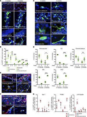 Mice were sacrificed and brains sectioned for immunohistochemical evidence of GFP+ donor cells. Original magnification, ×20 (CC and DG); ×40 (choroid plexus and perivascular region). n = 3 independent biological replicates. Data are presented as mean ± SEM. (B) Quantification of GFP+ cells in different brain regions evaluated at 2 and 8 weeks after irradiation showing a significant increase in the total number of GFP+ cells after 8 weeks in CC, DG, SVZ, and cortex. (C and D) Significant change in morphology of GFP+ cells over time with signs of cellular maturation and increase in branched morphology in various regions. Original magnification, ×40, except ×60 in DG and perivascular region, left-sided panel. (E–G) Phenotypical analysis of GFP+ cells demonstrate that the majority of cells colabel the microglial marker Iba-1 (see Figure 5C) and the monocyte-macrophage marker F4/80. Upper panel original magnification: ×20. Lower panel original magnification: ×40 (E). In addition, many GFP+ cells colabeled with B-III tubulin Upper panel original magnification: ×20. Lower panel original magnification: ×40 (F). (G) Quantification of GFP+ cells colabeling with Iba-1, F4/80, and B-III tubulin. Asterisks indicate a significant change relative to control. *P < 0.05; **P < 0.01; ***P < 0.001, 1-way ANOVA. n = 3 mice/group. Data are presented as mean ± SEM of biological replicates. |
Quantitative Spinal Cord MRI in Radiologically Isolated Syndrome
|
Publication: Neurol Neuroimmunol Neuroinflamm. 2018 Jan 17;5(2):e436. PMID: 29359174 | PDF Authors: Alcaide-Leon P, Cybulsky K, Sankar S, Casserly C, Leung G, Hohol M, Selchen D, Montalban X, Bharatha A, Oh J. Institution: Department of Surgery, St. Michael's Hospital, University of Toronto, ON, Canada. Abstract: OBJECTIVES: To assess whether quantitative spinal cord MRI (SC-MRI) measures, including atrophy, and diffusion tensor imaging (DTI) and magnetization transfer imaging metrics were different in radiologically isolated syndrome (RIS) vs healthy controls (HCs). METHODS: Twenty-four participants with RIS and 14 HCs underwent cervical SC-MRI on a 3T magnet. Manually segmented regions of interest circumscribing the spinal cord cross-sectional area (SC-CSA) between C3 and C4 were used to extract SC-CSA, fractional anisotropy, mean, perpendicular, and parallel diffusivity (MD, λ⊥, and λ||) and magnetization transfer ratio (MTR). Spinal cord (SC) lesions, SC gray matter (GM), and SC white matter (WM) areas were also manually segmented. Multivariable linear regression was performed to evaluate differences in SC-MRI measures in RIS vs HCs, while controlling for age and sex. RESULTS: In this cross-sectional study of participants with RIS, 71% had lesions in the cervical SC. Of quantitative SC-MRI metrics, spinal cord MTR showed a trend toward being lower in RIS vs HCs (p = 0.06), and there was already evidence of brain atrophy (p = 0.05). There were no significant differences in SC-DTI metrics, GM, WM, or CSA between RIS and HCs. CONCLUSION: The SC demonstrates minimal microstructural changes suggestive of demyelination and inflammation in RIS. These findings are in contrast to established MS and raise the possibility that the SC may play an important role in triggering clinical symptomatology in MS. Prospective follow-up of this cohort will provide additional insights into the role the SC plays in the complex sequence of events related to MS disease initiation and progression. "Manual segmentation of SC lesions was performed on the PSIR sequence between C1 and C7 using 3D Slicer by an experienced neuroradiologist." |
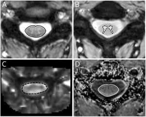 Quantitative spinal cord MRI maps and segmentations (A) Manual segmentation of the spinal cord cross-sectional area (black line) on high-resolution T2*-weighted, gradient-echo sequence with magnetization-transfer (MT) prepulse, (B) manual segmentation of spinal cord gray matter (black dashed line) on T2*-weighted, gradient-echo sequence without MT prepulse, (C) axial cross section of the map of fractional anisotropy with superimposed region of interest (black dashed line), and (D) axial cross section of the map of magnetization transfer ratio with superimposed region of interest (white line). |
Radiogenomic Analysis of Hypoxia Pathway is Predictive of Overall Survival in Glioblastoma
|
Publication: Sci Rep. 2018 Jan 8;8(1):7. PMID: 29311558 | PDF Authors: Beig N, Patel J, Prasanna P, Hill V, Gupta A, Correa R, Bera K, Singh S, Partovi S, Varadan V, Ahluwalia M, Madabhushi A, Tiwari P. Institution: Case Western Reserve University, Department of Biomedical Engineering, Cleveland, OH, USA. Abstract: Hypoxia, a characteristic trait of Glioblastoma (GBM), is known to cause resistance to chemo-radiation treatment and is linked with poor survival. There is hence an urgent need to non-invasively characterize tumor hypoxia to improve GBM management. We hypothesized that (a) radiomic texture descriptors can capture tumor heterogeneity manifested as a result of molecular variations in tumor hypoxia, on routine treatment naïve MRI, and (b) these imaging based texture surrogate markers of hypoxia can discriminate GBM patients as short-term (STS), mid-term (MTS), and long-term survivors (LTS). 115 studies (33 STS, 41 MTS, 41 LTS) with gadolinium-enhanced T1-weighted MRI (Gd-T1w) and T2-weighted (T2w) and FLAIR MRI protocols and the corresponding RNA sequences were obtained. After expert segmentation of necrotic, enhancing, and edematous/nonenhancing tumor regions for every study, 30 radiomic texture descriptors were extracted from every region across every MRI protocol. Using the expression profile of 21 hypoxia-associated genes, a hypoxia enrichment score (HES) was obtained for the training cohort of 85 cases. Mutual information score was used to identify a subset of radiomic features that were most informative of HES within 3-fold cross-validation to categorize studies as STS, MTS, and LTS. When validated on an additional cohort of 30 studies (11 STS, 9 MTS, 10 LTS), our results revealed that the most discriminative features of HES were also able to distinguish STS from LTS (p = 0.003). "The registration was performed using the General Registration (BRIANSFIT) module of 3D Slicer 4.5. Skull stripping was done using the skull-stripping module in 3D Slicer." |
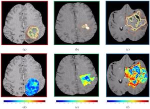 (a)–(c) show a 2D Gd-T1w MRI slice with expert-annotated necrosis (outlined in green), enhancing tumor (yellow) and edematous regions (brown) in 3 different GBM patients that exhibited low, medium, and high HES respectively. The corresponding Haralick feature map has been overlaid on the manually annotated tumor regions, for HESlow (d), HESmedium (e), and HEShigh (f). |
Paracrine Osteoprotegerin and β-catenin Stabilization Support Synovial Sarcomagenesis in Periosteal Cells
|
Publication: J Clin Invest. 2018 Jan 2;128(1):207-18. PMID: 29202462 | PDF Authors: Barrott JJ, Illum BE, Jin H, Hedberg ML, Wang Y, Grossmann A, Haldar M, Capecchi MR, Jones KB. Institution: Departments of Orthopaedics and Oncological Sciences, University of Utah, Salt Lake City, UT, USA. Abstract: Synovial sarcoma (SS) is an aggressive soft-tissue sarcoma that is often discovered during adolescence and young adulthood. Despite the name, synovial sarcoma does not typically arise from a synoviocyte but instead arises in close proximity to bones. Previous work demonstrated that mice expressing the characteristic SS18-SSX fusion oncogene in myogenic factor 5-expressing (Myf5-expressing) cells develop fully penetrant sarcomagenesis, suggesting skeletal muscle progenitor cell origin. However, Myf5 is not restricted to committed myoblasts in embryos but is also expressed in multipotent mesenchymal progenitors. Here, we demonstrated that human SS and mouse tumors arising from SS18-SSX expression in the embryonic, but not postnatal, Myf5 lineage share an anatomic location that is frequently adjacent to bone. Additionally, we showed that SS can originate from periosteal cells expressing SS18-SSX alone and from preosteoblasts expressing the fusion oncogene accompanied by the added stabilization of β-catenin, which is a common secondary change in SS. Expression and secretion of the osteoclastogenesis inhibitory factor osteoprotegerin enabled early growth of SS18-SSX2-transformed cells, indicating a paracrine link between the bone and synovial sarcomagenesis. These findings explain the skeletal contact frequently observed in human SS and may provide alternate means of enabling SS18-SSX-driven oncogenesis in cells as differentiated as preosteoblasts. Funding:
|
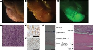 Expression of SS18-SSX2 in postnatal Prx1CreERT2 lineage induces synovial sarcomagenesis. "Reconstructed images were analyzed and visualized using 3D Slicer, v.4.6.2"" |
|
Publication: Int J Comput Assist Radiol Surg. 2018 Jan;13(1):115-24. PMID: 28718001 Authors: Doba N, Fukuda H, Numata K, Hao Y, Hara K, Nozaki A, Kondo M, Chuma M, Tanaka K, Takebayashi S, Koizumi N, Kobayashi A, Tokuda J, Maeda S. Institution: Gastroenterological Center, Yokohama City University Medical Center, Yokohama, Japan. Abstract: Purpose: Radiofrequency ablation for liver tumors (liver RFA) is widely performed under ultrasound guidance. However, discriminating between the tumor and the needle is often difficult because of cavitation caused by RFA-induced coagulation. An unclear ultrasound image can lead to complications and tumor residue. Therefore, image-guided navigation systems based on fiducial registration have been developed. Fiducial points are usually set on a patient's skin. But the use of internal fiducial points can improve the accuracy of navigation. In this study, a new device is introduced to use internal fiducial points using 2D US. Methods: 3D Slicer as the navigation software, Polaris Vicra as the position sensor, and two target tumors in a 3D abdominal phantom as puncture targets were used. Also, a new device that makes it possible to obtain tracking coordinates in the body was invented. First, two-dimensional reslice images from the CT images using 3D Slicer were built. A virtual needle was displayed on the two-dimensional reslice image, reflecting the movement of the actual needle after fiducial registration. A phantom experiment using three sets of fiducial point configurations: one conventional case using only surface points, and two cases in which the center of the target tumor was selected as a fiducial point was performed. For each configuration, one surgeon punctured each target tumor ten times under guidance from the 3D Slicer display. Finally, a statistical analysis examining the puncture error was performed. Results: The puncture error for each target tumor decreased significantly when the center of the target tumor was included as one of the fiducial points, compared with when only surface points were used. Conclusion: This study introduces a new device to use internal fiducial points and suggests that the accuracy of image-guided navigation systems for liver RFA can be improved by using the new device. |
|
Publication: Acta Neuropathol. 2018 Jan;135(1):95-113. PMID: 29116375 | PDF Authors: von Jonquieres G, Spencer ZHT, Rowlands BD, Klugmann CB, Bongers A, Harasta AE, Parley KE, Cederholm J, Teahan O, Pickford R, Delerue F, Ittner LM, Fröhlich D, McLean CA, Don AS, Schneider M, Housley GD, Rae CD, Klugmann M Institution: Translational Neuroscience Facility and Department of Physiology, School of Medical Sciences, UNSW Sydney, Sydney, Australia. Abstract: N-Acetylaspartate (NAA) is the second most abundant organic metabolite in the brain, but its physiological significance remains enigmatic. Toxic NAA accumulation appears to be the key factor for neurological decline in Canavan disease-a fatal neurometabolic disorder caused by deficiency in the NAA-degrading enzyme aspartoacylase. To date clinical outcome of gene replacement therapy for this spongiform leukodystrophy has not met expectations. To identify the target tissue and cells for maximum anticipated treatment benefit, we employed comprehensive phenotyping of novel mouse models to assess cell type-specific consequences of NAA depletion or elevation. We show that NAA-deficiency causes neurological deficits affecting unconscious defensive reactions aimed at protecting the body from external threat. This finding suggests, while NAA reduction is pivotal to treat Canavan disease, abrogating NAA synthesis should be avoided. At the other end of the spectrum, while predicting pathological severity in Canavan disease mice, increased brain NAA levels are not neurotoxic per se. In fact, in transgenic mice overexpressing the NAA synthesising enzyme Nat8l in neurons, supra-physiological NAA levels were uncoupled from neurological deficits. In contrast, elimination of aspartoacylase expression exclusively in oligodendrocytes elicited Canavan disease like pathology. Although conditional aspartoacylase deletion in oligodendrocytes abolished expression in the entire CNS, the remaining aspartoacylase in peripheral organs was sufficient to lower NAA levels, delay disease onset and ameliorate histopathology. However, comparable endpoints of the conditional and complete aspartoacylase knockout indicate that optimal Canavan disease gene replacement therapies should restore aspartoacylase expression in oligodendrocytes. On the basis of these findings we executed an ASPA gene replacement therapy targeting oligodendrocytes in Canavan disease mice resulting in reversal of pre-existing CNS pathology and lasting neurological benefits. This finding signifies the first successful post-symptomatic treatment of a white matter disorder using an adeno-associated virus vector tailored towards oligodendroglial-restricted transgene expression. "Whole brain and ventricle structures were segmented using thresholding and delineation methods provided by the 3D Slicer package." |
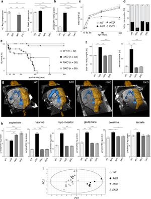 Pathological consequences of manipulation of NAA metabolism. a qRT-PCR analysis of Aspa and Nat8l mRNA expression in CNS tissue from DKO and the parental single gene mutants (n = 3; expression relative to Hprt). Segmentation of images was performed in 3D Slicer outlining brain surface (yellow) and ventricles (blue). |
Go to 2020 :: 2019 :: 2018 :: 2017 :: 2016 :: 2015 :: 2014-2011:: 2010-2005

