Documentation/Nightly/Modules/SegmentationSmoothing
|
For the latest Slicer documentation, visit the read-the-docs. |
Introduction and Acknowledgements
|
Module Description
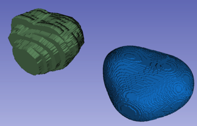
| This module can be used to generate a smooth surface segmentation for datasets that were segmented on images with large slice thickness, leading to "staircase" effect. This module was motivated by the need to generate smooth segmentations for prostate MRI images, which typically have high in-plane resolution (0.5mm) relative to the slice thickness (3mm).
The module operates by resampling the input to isotropic voxel resolution and applying Gaussian smoothing to the resampled image. Note that the resulting image will have larger size than the input. |
Use Cases
- MRI-ultrasound fusion biopsy of the prostate (primary)
- therapy planning
- treatment response assessment
Tutorials
None at this time ... stay tuned!
Parameters:
- IO: Input/output parameters
- Input label (inputImageName): Input segmentation label
- Output label (outputImageName): Smoothed segmentation label
- Label to process (labelNumber): Label to smooth in the input label image. Default value of -1 will threshold the input image between 1 and 255 and will apply smoothing to the result.
List of parameters generated transforming this XML file using this XSL file. To update the URL of the XML file, edit this page.
Similar Modules
References
[1] Fedorov A, Khallaghi S, Antonio Sánchez C, Lasso A, Fels S, Tuncali K, Neubauer Sugar E, Kapur T, Zhang C, Wells W, Nguyen PL, Abolmaesumi P, Tempany C. (2015) Open-source image registration for MRI–TRUS fusion-guided prostate interventions. Int J CARS: 1–10. Available: http://link.springer.com/article/10.1007/s11548-015-1180-7.
[2] Fedorov A, Nguyen PL, Tuncali K, Tempany C. (2015). Annotated MRI and ultrasound volume images of the prostate. Zenodo. http://doi.org/10.5281/zenodo.16396


