Home < Documentation < 4.10 < Modules < DistanceMapBasedRegistration
Introduction and Acknowledgements
|

|
Extension: SlicerProstate
Acknowledgments:
This work supported in part the National Institutes of Health, National Cancer Institute through the following grants:
- Quantitative MRI of prostate cancer as a biomarker and guide for treatment, Quantitative Imaging Network (U01 CA151261, PI Fennessy)
- Enabling technologies for MRI-guided prostate interventions (R01 CA111288, PI Tempany)
- The National Center for Image-Guided Therapy (P41 EB015898, PI Tempany)
- Quantitative Image Informatics for Cancer Research (QIICR) (U24 CA180918, PIs Kikinis and Fedorov).
Authors: Andrey Fedorov (SPL), Andras Lasso (Queen's University)
Contact: Andrey Fedorov, <email>fedorov@bwh.harvard.edu</email>
License: Slicer License
|
| National Center for Image Guided Therapy (NCIGT)
|
| Quantitative Image Informatics for Cancer Research
|
| Surgical Planning Laboratory (SPL)
|
|
|
Module Description
| This module was originally developed to enable registration between segmented MRI and transrectal ultrasound (TRUS) images of the prostate. The registration approach is based on hierarchical deformable registration of the smoothed distance maps of the segmentation masks (see details in the publication provided in the references section), and thus is not modality- or organ-specific, and can be applied in other applications.
|
 Example visualization of MRI/TRUS registration result obtained using this module. Yellow outline is the contour of the manual segmentation of the prostate gland in TRUS, over the registered MRI image of the same prostate. |
Use Cases
- MRI-ultrasound fusion biopsy of the prostate (primary)
- therapy planning
- treatment response assessment
Tutorials
None at this time ... stay tuned!
Panels and their use
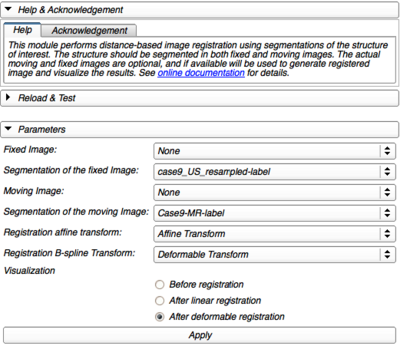
- Fixed image (optional): image that should be used as reference in registration
- Segmentation of the fixed image
- Moving image (optional): image to be registered
- Segmentation of the moving image
- Registration affine transform: transform to store the result of affine registration
- Registration deformable transform: transform to store the result of deformable registration
- Visualization: initializes slice viewers to show overlay of the fixed and moving images (before or after registration), outline of the fixed image segmentation and the registration transform grid and visualization of the fixed and moving segmentation label surfaces in 3d view
|
|
Similar Modules
Slicer Registration modules and extensions.
References
[1] Fedorov A, Khallaghi S, Antonio Sánchez C, Lasso A, Fels S, Tuncali K, Neubauer Sugar E, Kapur T, Zhang C, Wells W, Nguyen PL, Abolmaesumi P, Tempany C. (2015) Open-source image registration for MRI–TRUS fusion-guided prostate interventions. Int J CARS: 1–10. Available: http://link.springer.com/article/10.1007/s11548-015-1180-7.
[2] Fedorov A, Nguyen PL, Tuncali K, Tempany C. (2015). Annotated MRI and ultrasound volume images of the prostate. Zenodo. http://doi.org/10.5281/zenodo.16396
Information for Developers


