White Matter Exploration Tutorial
From Slicer Wiki
Home < White Matter Exploration Tutorial
White Matter Exploration for Neurosurgical Planning
Authors: Sonia Pujol, Ph.D., Ron Kikinis, M.D.
Surgical Planning Laboratory, Brigham and Women's Hospital, Harvard Medical School, Boston, MA
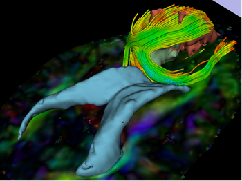
- Summary: Diffusion Tensor Imaging (DTI) Tractography has the potential to bring valuable spatial information on tumor infiltration and tract displacement for neurosurgical planning of tumor resection. This tutorial walks the user through an integrated workflow for segmenting the solid and cystic part of a tumor in a Glioblastoma multiforme (GBM) case, and reconstructing the fibers surrounding the pathology using streamline tractography. The pipeline includes an interactive 3D exploration of the white matter architecture on the ipsilateral and contralateral side using "tractography on the fly".
- Category: Workflow
- Dataset:The tutorial dataset contains a baseline volume and tensor volume in Nrrd file format (size: 18 MB).
- Slicer version: Slicer3.6.3 release (March 2011)
- Audience: Clinicians, Clinical researchers, scientists
- Estimated completion time: 1 hour
