Documentation/4.6/Modules/SlicerModuleCMRToolkitWizard
From Slicer Wiki
Home < Documentation < 4.6 < Modules < SlicerModuleCMRToolkitWizard
|
For the latest Slicer documentation, visit the read-the-docs. |
Introduction and Acknowledgements
|
Author: Umme Salma Bengali, CARMA Center, University of Utah | |
|
|
Module Description
This wizard module takes the user through the steps involved in analyzing a cardiac LGE-MRI image for scar enhancement. The wizard uses modules from the Cardiac MRI Toolkit extension as well as core Slicer modules. Test data or other data can be used to run the wizard.
Use Cases
This module is meant to be used for cardiac LGE-MRI scans.
Tutorials
[[Media:]]
Panels and their use
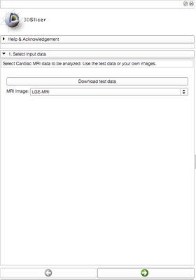
- Select the input cardiac MRI image.
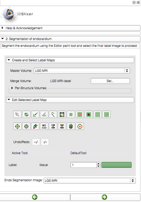
- Manually segment the left atrial (LA) endocardium region.
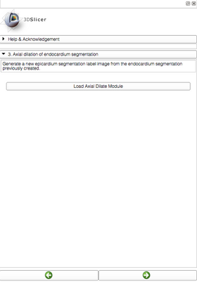
- Axially dilate the endocardium segmentation to obtain the epicardium region.
- Subtract the endocardium segmentation from the epicardium segmentation to obtain the wall region.
- Remove the pulmonary veins (PVs) from the wall segmentation.
- Cut off the PVs from the endocardium segmentation created in step 2.
- Create the isosurface of the endocardium segmentation without the PVs.
- Run the automatic scar module to view regions of scar enhancement in the MRI. Create the isosurface of the scar to view it overlaid on the endocardium isosurface.
Similar Modules
N/A
References
N/A
Information for Developers
The source code for this module can be found at the CARMA GitHub repository.


