Documentation/Nightly/Extensions/SlicerPathology

From Slicer Wiki
Revision as of 13:43, 13 May 2016 by Ebremer (talk | contribs)
Jump to: navigation, search
Home < Documentation < Nightly < Extensions < SlicerPathology


For the latest Slicer documentation, visit the read-the-docs.


Introduction and Acknowledgements

Extension: SlicerPathology
Acknowledgments: This work was supported by via the NIH-National Cancer Institute Grant U24 CA180918, as well as, U24 CA180918 Quantitative Image Informatics for Cancer Research (QIICR), http://qiicr.org, PIs Ron Kikinis and Andrey Fedorov, Brigham and Women's Hospital.
Author: Erich Bremer

Contributor 1: Yi Gao
Contributor 2: Nicole Aucoin (SPL)
Contributor 3: Andrey Fedorov (SPL)
Contributor 4: Jean-Christophe Fillion-Robin (Kitware)
Contact: Erich Bremer, <email>erich.bremer@stonybrook.edu</email>

Module Description


Use Cases


Tutorials

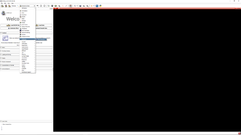
Step 1 - Go to the SlicerPathology Extension.
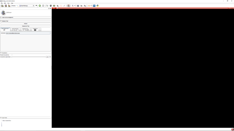
Step 2 - Click the user information tab. Enter your email address. This is used for identification of you in the file data files.
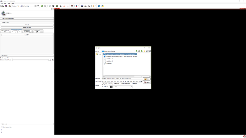
Step 3 - Click "Load data" to select an image.
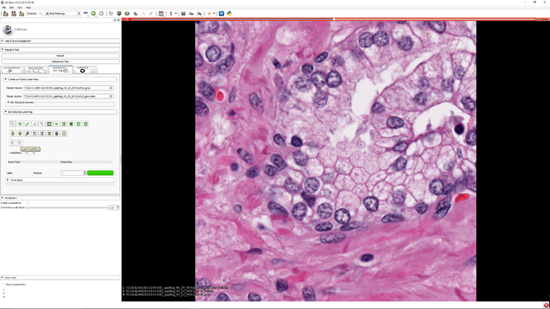
Step 4 - Click the button Quick TCGA Effect button to activate the effect.
Step 5 - Click "Start TCGA Segmenter and adjust the five segmentation algorithm parameters as needed.
Step 6 - you can select a subregion to speed up the parameter tuning process. Press "Y" to execute the segmenter on the subregion (if it was selected) or the whole image
Step 7 - you can click "Clear Selection" to clear the selected sub-region so that the segmenter operates on the whole image.
Step 8 - "Click the "Submission" tab so that you can save your label masks and related meta data. (meta data will be stored as JSON)

Panels and their use


References

Information for Developers