Documentation/Nightly/Modules/LASegmenter
|
For the latest Slicer documentation, visit the read-the-docs. |
Introduction and Acknowledgements
|
This work is part of the National Alliance for Medical Image Computing (NA-MIC), funded by the National Institutes of Health through the NIH Roadmap for Medical Research, Grant U54 EB005149. Information on NA-MIC can be obtained from the NA-MIC website. Contributor: Liangjia Zhu, Stony Brook University | |||||||||
|
Module Description
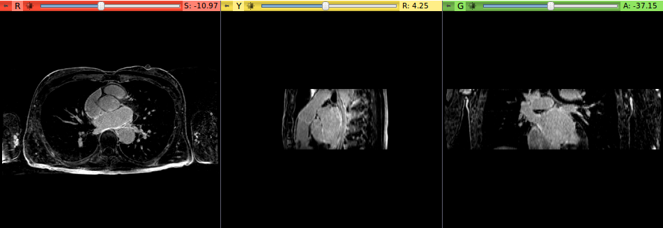
This module performs the segmentation of the left atrium from MR images. It is an implementation of a region-growing-based method with a shape prior. The module starts from a manually generated seed region inside the left atrium. Then, the growing process gradually captures the whole left atrium region.
Use Cases
This module is specifically designed for the segmentation of the left atrium from MR images.
Tutorials
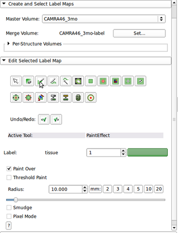
- Select Editor module
- Choose Master Volume and Merge Volume (label map to be used for the left atrium seed region)
- Select PaintEffect and adjust Radius (in mm) so that the painted seed region (a circle) is completely inside the left atrium
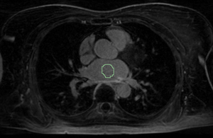
- Go to the Axial view, choose a slice that is approximately to the center of the left atrium. Then, draw a circle that lies to the center of the left atrium on the slice
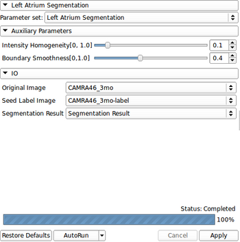
- Go to Segmentation->Left Atrium Segmentation module
- Select Original Image, Seed Label Image (the one obtained from the Editor module), and select/create Segmentation Results
- Adjust parameters if needed
- Press Apply button and it takes several minutes to process one volume
Panels and their use
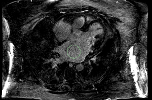
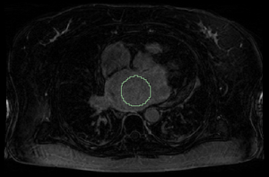
A properly set seed region is required to start the segmentation process. In principle, the seed region needs to be set closely to the center of the left atrium and covers the most of left atrium region on the selected image slice. The radius of a seed region is determined by the size of the left atrium. A small radius may cause the segmentation terminates before reaching the left atrium boundary. A large radius may lead to oversegmentation. Examples of good seed regions with various radius from small to large are shown below.
From left to right: seed radii are 10, 18, and 24 mm, respectively.
Similar Modules
References
L. Zhu, Y. Gao, A. Yezzi, R. MacLeod, J. Cates, A. Tannenbaum. Automatic Segmentation of the Left Atrium from MRI Images Using Salient Feature and Contour Evolution, IEEE Engineering in Medicine and Biology Conference(EMBC), 2012.
Information for Developers
| Section under construction. |


