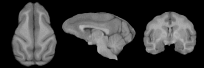
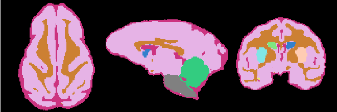
EMSegmenter-Tasks:Non-Human-Primate
Return to EMSegmenter Task Overview Page
Description
Non Human Primate pipeline facilitates segmentation of single-channel (T1w) MRI of the vervet brain. The current segmentation pipeline task operates on the skull-stripped brain MRI. In our experience, skull-stripping of NHP brain can be performed by registration of a subject with manually defined ICC to the subject being analyzed. We recommend using BRAINSFit module for registering ICC template to the subject. In order to improve the robustness of the registration process we recommend prior bias correction using N4ITKBiasFieldCorrection module. The resulting ICC may need to be manually adjusted using Editor module. In the future, we plan to substitute this registration-based skull-stripping approach with the SPECTRE Slicer plugin currently under development.
The current pipeline consist of the following steps:
- Step 1: Register the atlas to the MRI scan via BRAINSFit (Johnson et al 2007)
- Step 2: Compute the intensity distributions for each structure
Compute intensity distribution (mean and variance) for each label by automatically sampling from the MR scan. The sampling for a specific label is constrained to the region that consists of voxels with high probability (top 95%) of being assigned to the label according to the aligned atlas.
- Step 3: Automatically segment the MRI scan into the structures of interest using EM Algorithm (Pohl et al 2007)
Anatomical Tree
- Root
- Head
- intracranial cavity (ICC)
- White matter + Gray matter + CSF class (WGC)
- grey matter (GM)
- left Subcortical Gray Matter (lSGM)
- left caudate (lCaudate)
- left putamen (lPutamen)
- left hippocampus (lHippocampus)
- right Subcortical Gray Matter (rSGM)
- right caudate (rCaudate)
- right putamen (rPutamen)
- right hippocampus (rHippocampus)
- Cortical Gray Matter (CGM)
- left Subcortical Gray Matter (lSGM)
- white matter (WM)
- cerebrospinal fluid (CSF)
- grey matter (GM)
- Brainstem
- Cerebellum
- White matter + Gray matter + CSF class (WGC)
- background (BG)
- intracranial cavity (ICC)
- Head
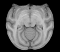
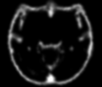
Atlas
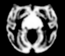
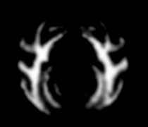
Image Dimension = 256 x 256 x 252
Image Spacing = 0.4688 x 0.4688 x 0.50002
|
|
|
|
| Template (T1) | CSF | GM | WM |
Result
Collaborators
Andriy Fedorov (BWH)
Citations
- Tustison NJ, Avants BB, Cook PA, Zheng Y, Egan A, Yushkevich PA, Gee JC N4ITK: Improved N3 Bias Correction, IEEE Trans Med Imag, 2010
- Pohl K, Bouix S, Nakamura M, Rohlfing T, McCarley R, Kikinis R, Grimson W, Shenton M, Wells W. A Hierarchical Algorithm for MR Brain Image Parcellation. IEEE Transactions on Medical Imaging. 2007 Sept;26(9):1201-1212.
- S. Warfield, J. Rexilius, P. Huppi, T. Inder, E. Miller, W. Wells, G. Zientara, F. Jolesz, and R. Kikinis, “A binary entropy measure to assess nonrigid registration algorithms,” in MICCAI, LNCS, pp. 266–274, Springer, October 2001.
- Johnson H.J., Harris G., Williams K. BRAINSFit: Mutual Information Registrations of Whole-Brain 3D Images, Using the Insight Toolkit, The Insight Journal, July 2007
