EMSegmenter-Tasks:MRI-Human-Brain
From Slicer Wiki
Home < EMSegmenter-Tasks:MRI-Human-Brain
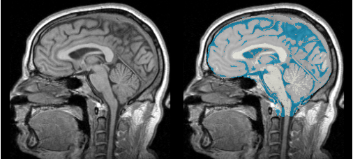
MRI Human Brain
Description
Automatic segmentation of t1w-MRI brain scans into the major tissue classes (gray matter, white matter, csf). The pipeline only works correctly if the MRI brain scan shows part of the skull and neck. The pipeline performs the following steps:
Anatomical Tree
- root
- background (BG)
- air (AIR)
- skull (skull)
- intracranial cavity (ICC)
- white matter (WM)
- grey matter (GM)
- cerebrospinal fluid (CSF)
- background (BG)
Atlas
Image Dimension = 256 x 256 x 124
Image Spacing = 0.9375 x 0.9375 x 0.1.5
Pre-Processing
- 1 step: image inhomogeneity correction N4ITKBiasFieldCorrection
- 2 step: atlas-to-target registration BRAINSFit
- 3 step: automatic mean/covariance calculation. (TODO Kilian)
