EMSegmenter-Tasks:MRI-Human-Brain
From Slicer Wiki
Home < EMSegmenter-Tasks:MRI-Human-Brain
MRI Human Brain
Description
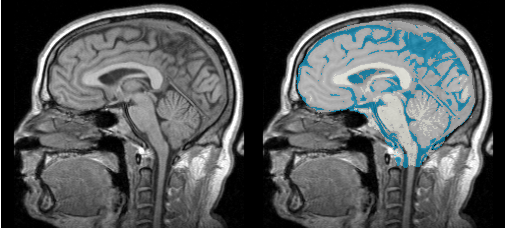
Single channel automatic segmentation of t1w-MRI brain scans into the major tissue classes (gray matter, white matter, csf). The task can only be applied to t1w brain scan showing parts of the skull and neck. The pipeline consist of the following steps:
- Step 1: Perform image inhomogeneity correction of the MRI scan via N4ITKBiasFieldCorrection (Tustison et al 2010)
- Step 2: Register the atlas to the MRI scan via BRAINSFit (ask Hans for citation)
- Step 3: Compute the intensity distributions for each structure
- Step 4: Automatically segment the MRI scan into the structures of interest using the EM Algorithm (Pohl et al 2007)
Anatomical Tree
- root
- background (BG)
- air (AIR)
- skull (skull)
- intracranial cavity (ICC)
- white matter (WM)
- grey matter (GM)
- cerebrospinal fluid (CSF)
- background (BG)
Atlas
Image Dimension = 256 x 256 x 124
Image Spacing = 0.9375 x 0.9375 x 0.1.5
Pre-Processing
Result
Citations
- Tustison NJ, Avants BB, Cook PA, Zheng Y, Egan A, Yushkevich PA, Gee JC N4ITK: Improved N3 Bias Correction, IEEE Trans Med Imag, 2010
- Pohl K, Bouix S, Nakamura M, Rohlfing T, McCarley R, Kikinis R, Grimson W, Shenton M, Wells W. A Hierarchical Algorithm for MR Brain Image Parcellation. IEEE Transactions on Medical Imaging. 2007 Sept;26(9):1201-1212.
