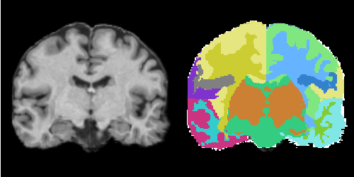
EMSegmenter-Tasks:MRI-Human-Brain-Parcellation
From Slicer Wiki
Home < EMSegmenter-Tasks:MRI-Human-Brain-Parcellation
Description
MRI Human Head pipeline for a finer-grained parcellation
Anatomical Tree
The anatomical tree represents the structures to be segmented. Node labels displayed below contain a human readable structure name and in parentheses the internally used structure name.
- Root
- background (BG)
- grey matter (GM)
- left temporal grey matter (LTGM)
- left temporal grey matter - region 1 (LTGM1)
- left temporal grey matter - region 2 (LTGM2)
- left temporal grey matter - region 3 (LTGM3)
- left temporal grey matter - region 4 (LTGM4)
- right temporal grey matter (RTGM)
- right temporal grey matter - region 1 (RTGM1)
- right temporal grey matter - region 2 (RTGM2)
- right temporal grey matter - region 3 (RTGM3)
- right temporal grey matter - region 4 (RTGM4)
- TODO (SUBGM)
- left temporal grey matter (LTGM)
- white matter (WM)
- left temporal white matter (LTWM)
- left temporal white matter - region 1 (LTWM1)
- left temporal white matter - region 2 (LTWM2)
- left temporal white matter - region 3 (LTWM3)
- left temporal white matter - region 4 (LTWM4)
- right temporal white matter (RTWM)
- right temporal white matter - region 1 (RTWM1)
- right temporal white matter - region 2 (RTWM2)
- right temporal white matter - region 3 (RTWM3)
- right temporal white matter - region 4 (RTWM4)
- TODO (SUBWM)
- left temporal white matter (LTWM)
- cerebrospinal fluid (CSF)
Atlas
Image Dimension = 256 x 256 x 220
Image Spacing = 0.9375 x 0.9375 x 0.9375
Pre-Processing
Pre procssing includes atlas-to-target registration, image inhomogeneity correction, and automatic mean/covariance calculation.
Result
Collaborators
Padmapriya Srinivasan and Sylvain Bouix (PNL-BWH)
