Documentation/Nightly/Modules/PETTumorSegmentationEffect
|
For the latest Slicer documentation, visit the read-the-docs. |
Introduction and Acknowledgements
|
Acknowledgments:
This work is funded in part by Quantitative Imaging to Assess Response in Cancer Therapy Trials NIH grant U01-CA140206 and Quantitative Image Informatics for Cancer Research (QIICR) NIH grant U24 CA180918. License: Slicer License | |||||||
|
Module Description
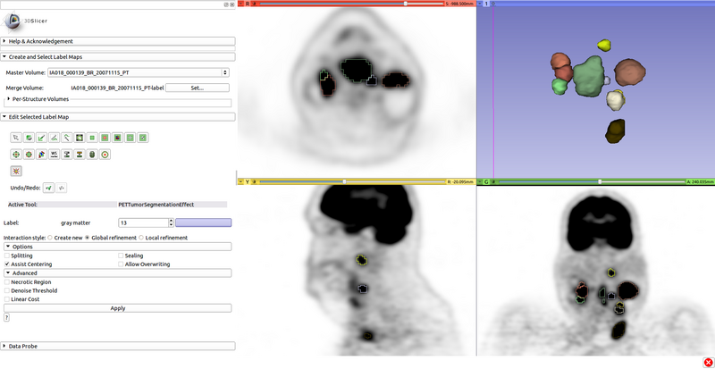
Editor Effect for segmentation of tumors and hot lymph nodes in PET scans.
Tutorials
Click on a lesion (tumor or hot lymph node) in a PET scan to segment it. Depending on refinement settings, click again to refine globally and/or locally. Options may help deal with cases such as segmenting individual lesions in a chain.
| Section under construction. |
References
- Markus L. van Tol (2014): A Graph-Based Method for Segmentation of Tumors and Lymph Nodes in Volumetric PET Images, The University of Iowa, 2014
Information for Developers
| Section under construction. |


