Difference between revisions of "Modules:MRIBiasFieldCorrection-Documentation-3.5"
From Slicer Wiki
Sylvainjaume (talk | contribs) (→Authors, Collaborators & Contact: add contact email) |
Sylvainjaume (talk | contribs) (→MRI Bias Field Correction: add screenshots of MRIBiasFieldCorrection module applied on the SPL 3D Brain Atlas) |
||
| Line 6: | Line 6: | ||
===MRI Bias Field Correction=== | ===MRI Bias Field Correction=== | ||
Filtering:MRIBiasFieldCorrection | Filtering:MRIBiasFieldCorrection | ||
| + | |||
| + | {| | ||
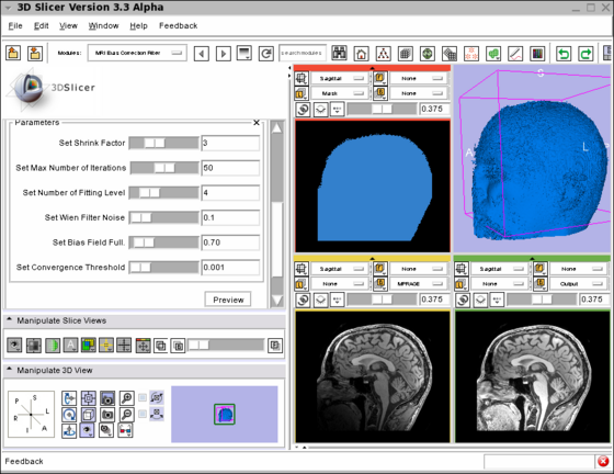
| + | |[[Image:MRI_Bias_Field_Correction_Slicer3.png|thumb|560px|Screenshot of the MRIBiasFieldCorrection module in Slicer3. | ||
| + | The screenshot illustrates the application of the MRI Bias Field Correction on the SPL Atlas. | ||
| + | The top left window shows the mask used to defined the ROI where the correction will be applied. | ||
| + | The top right window shows the rendering of the 3D model created using the mask. | ||
| + | The bottom left window shows one slice in the input image. | ||
| + | Note the intensity inhomogeneity from the bottom left corner (dark) to the top right corner (bright). | ||
| + | The bottom right window shows the output image after the application of the MRI Bias Field Correction module. | ||
| + | The parameters used for the correction are shown on the left panel of the Slicer3 interface.]] | ||
| + | |} | ||
| + | |||
| + | {| | ||
| + | |[[Image:MRI_Bias_Field_Correction_Slicer3_close_up.png|thumb|560px|Close-up of the above screenshot showing the image before and after correction. | ||
| + | Note that the intensity inhomogeneity visible in the input image (left) has been corrected in the output image (right).]] | ||
| + | |} | ||
{| | {| | ||
Revision as of 21:36, 20 June 2009
Home < Modules:MRIBiasFieldCorrection-Documentation-3.5Return to Slicer 3.5 Documentation
MRI Bias Field Correction
Filtering:MRIBiasFieldCorrection
General Information
Module Type & Category
Type: Interactive or CLI
Category: Filtering
Authors, Collaborators & Contact
- Nicolas Rannou: Harvard Medical School, Brigham and Women's Hospital
- Sylvain Jaume: MIT Computer Science and Artificial Intelligence Laboratory
- Contact: <nrannou at bwh.harvard.edu>, <sylvain at csail.mit.edu>
Module Description
The module filters the image to remove the intensity inhomogeneity due to the MRI image acquisition.
Usage
- Load the input dataset (Add Volume)
- Go to the menu Modules > Filtering > MRI Bias Field Correction
- Select the input dataset
- Modify the number of iterations (large values make the processing longer)
- Modify the fitting levels
- Click on Apply (processing of large images take in the order of minutes)
- Select the 'field' checkbox if you want to visualize the field
Examples, Use Cases & Tutorials
- Note use cases for which this module is especially appropriate, and/or link to examples. (to be defined)
- Link to examples of the module's use (to be defined)
- Link to any existing tutorials (to be defined)
Quick Tour of Features and Use
List all the panels in your interface, their features, what they mean, and how to use them. For instance:
- Input panel:
- Parameters panel:
- Output panel:
- Viewing panel:
Development
Dependencies
No other modules or packages are required to use this module.
Known bugs
Follow this link to the Slicer3 bug tracker.
Usability issues
Follow this link to the Slicer3 bug tracker. Please select the usability issue category when browsing or contributing.
Source code & documentation
More Information
Acknowledgment
This work has been supported by NA-MIC Algorithms.
References
- A Nonparametric Method for Automatic Correction of Intensity Nonuniformity in MRI Data, J.G. Sled, A.P. Zijdenbos, and A.C. Evans, IEEE Transactions on Medical Imaging, 17(1):87–97, Feb 1998.
- Parametric Estimate of Intensity Inhomogeneities Applied to MRI, M. Styner, C. Brechbhler, G. Szekely, and G. Gerig, IEEE Transactions on Medical Imaging, 19(3):153–165, Mar 2000.
- N4ITK: Nick's N3 ITK Implementation For MRI Bias Field Correction, Tustison N., Gee J., Insight Journal, 2009.


