Difference between revisions of "Stanford Simbios group"
| Line 15: | Line 15: | ||
==Atlas Generation from Input MR Images== | ==Atlas Generation from Input MR Images== | ||
===Model Generation from Input MR Images=== | ===Model Generation from Input MR Images=== | ||
| − | Pre-Segmented models for femur, patella and tibia are obtained for a patient (in .stl format) | + | Pre-Segmented models for femur, patella and tibia are obtained for a patient (in .stl format). |
[[Image:All_Three.JPG|left|thumb| Pre-Segmented Femur/Patella/Tibia Model]] | [[Image:All_Three.JPG|left|thumb| Pre-Segmented Femur/Patella/Tibia Model]] | ||
| Line 33: | Line 33: | ||
We are now in the process of trying to register new patient image to the existing patient image. If we register the images, we can get the transform which can be used to register the existing atlas (label map) to the new patient. | We are now in the process of trying to register new patient image to the existing patient image. If we register the images, we can get the transform which can be used to register the existing atlas (label map) to the new patient. | ||
| − | We | + | We think that Pipelined Bspline Registration may give promising results for our dataset. Below are some of the results. Here we consider 64_MRI as the fixed image and 58_MRI as the moving image and used '''Pipelined BSpline Image registration method''' for registration. Also we gave one point as landmark point. |
<gallery widths="400px" perrow="6"> | <gallery widths="400px" perrow="6"> | ||
Image:58_MRI.jpg | Image:58_MRI.jpg | ||
| Line 40: | Line 40: | ||
</gallery> | </gallery> | ||
| − | + | We also tried affine registration for the above mentioned patients with 64 as fixed MRI and 58 as moving MRI and one landmark point. The scene file for this is | |
| + | [[Image:SlicerScene1 64 Fixed Affine Registration 58 moving.mrml|left|64_Affine_Registered_Scene]] | ||
| + | <gallery widths="400px" perrow="6"> | ||
| + | Image:64 Affine Registration.JPG | ||
| + | </gallery> | ||
===Multi Image Registration=== | ===Multi Image Registration=== | ||
Revision as of 23:35, 7 May 2009
Home < Stanford Simbios group
Contents
Aim
To develop a generic semi-automatic segmentation toolkit to convert images of musculoskeletal structures to 3D models.
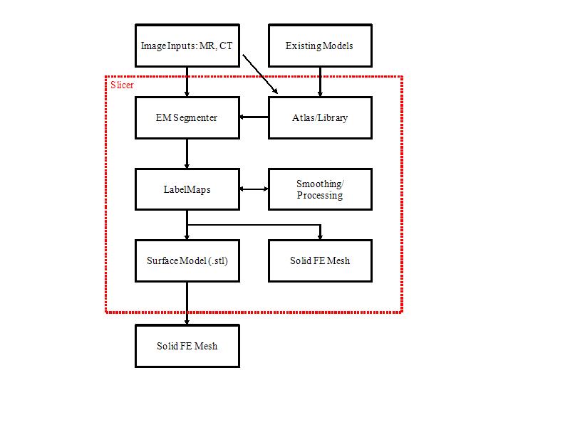
Process Flowchart
Progress
Atlas Generation from Input MR Images
Model Generation from Input MR Images
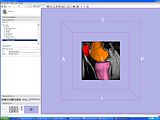
Pre-Segmented models for femur, patella and tibia are obtained for a patient (in .stl format).
Create a filled label map using PolyDataToFilledLabelMap module in slicer
The models generated in the above step are closed but hollow. But EM Segmentation requires the atlas given to be in closed filled format. Hence we converted the hollow closed models into filled label volumes using the module PolyDataToFilledLabelMap module in Slicer. The output label map of this module is of datatype unsigned char and is converted to short datatype.
EM Segmentation based on the atlas
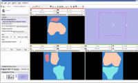
The output label map in the above is given as input atlas in the EM Segmentation step. As an initial step we ran EM Segmentation on the same patient. The EM Segmented output is given in the below figure.
Register Images
Register Images Module in Slicer
We are now in the process of trying to register new patient image to the existing patient image. If we register the images, we can get the transform which can be used to register the existing atlas (label map) to the new patient.
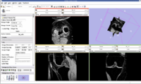
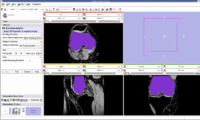
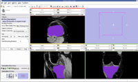
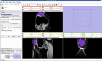
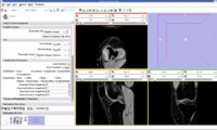
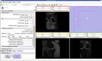
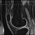
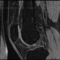
We think that Pipelined Bspline Registration may give promising results for our dataset. Below are some of the results. Here we consider 64_MRI as the fixed image and 58_MRI as the moving image and used Pipelined BSpline Image registration method for registration. Also we gave one point as landmark point.
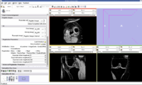
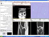
We also tried affine registration for the above mentioned patients with 64 as fixed MRI and 58 as moving MRI and one landmark point. The scene file for this is File:SlicerScene1 64 Fixed Affine Registration 58 moving.mrml
Multi Image Registration
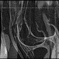
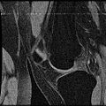
We have also tried the approach of Non-rigid Groupwise Registration using Bspline Deformation Model by Serdar K. Balci, Polina Golland and William M. Wells. In this approach, we try to give one slice each of two different patients we need to register as input. Here are some of the results of their approach.
- Slice 1:
- Slice 2:
- Mean Slice of above two :
- Slice 1:
- Slice 2:
- Mean Slice of above two :
Datasets
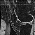
Below are the datasets that we used in our experiments. All the images are in standard dicom format.
