Difference between revisions of "Documentation/Nightly/Modules/LSContrastEnhancer"
From Slicer Wiki
Acsenrafilho (talk | contribs) (Added wiki page for LSContrastEnhancer module) |
Acsenrafilho (talk | contribs) m |
||
| Line 37: | Line 37: | ||
{{documentation/{{documentation/version}}/module-section|Panels and their use}} | {{documentation/{{documentation/version}}/module-section|Panels and their use}} | ||
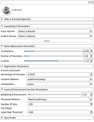
| − | [[Image: | + | [[Image:lscontrastenhancer_gui.png|thumb|380px|User Interface]] |
IO: | IO: | ||
*Input Volume | *Input Volume | ||
Revision as of 14:54, 9 December 2016
Home < Documentation < Nightly < Modules < LSContrastEnhancer
|
For the latest Slicer documentation, visit the read-the-docs. |
Introduction and Acknowledgements
|
Extension: LesionSpotlight | |||||||
|
Module Description
This module offer a simple application ...
Use Cases
- Use Case 1:
- Something.
- LSSegmenter FLAIR.png
Input T2-FLAIR image with multiple sclerosis lesions
- LSSegmenter Label.png
Lesion map resulted from LSSegmenter module
Panels and their use
IO:
- Input Volume
- Select the input image
- Output Volume
- Set the output image file which the filters should place the final result
... Parameters:
- Conductance
- The conductance regulates the diffusion intensity in the neighbourhood area. Choose a higher conductance if the input image has strong noise seem in the whole image space.
- See the reference paper[1] to choose the appropriate q value (at moment, only tested in MRI T1 and T2 weighted images).
Similar Modules
References
- paper
Information for Developers
| Section under construction. |
- ↑ Da S Senra Filho, A. C., Garrido Salmon, C. E., & Murta Junior, L. O. (2015). Anomalous diffusion process applied to magnetic resonance image enhancement. Physics in Medicine and Biology, 60(6), 2355–2373. doi:10.1088/0031-9155/60/6/2355


