Difference between revisions of "Documentation/Nightly/Modules/EpileptogenicFocusDetection"
From Slicer Wiki
| Line 8: | Line 8: | ||
{{documentation/{{documentation/version}}/module-introduction-start|{{documentation/modulename}}}} | {{documentation/{{documentation/version}}/module-introduction-start|{{documentation/modulename}}}} | ||
{{documentation/{{documentation/version}}/module-introduction-row}} | {{documentation/{{documentation/version}}/module-introduction-row}} | ||
| − | This work | + | This work was supported by Comision Sectorial de Investigacion Cientıfica (CSIC, Universidad de la Republica, Uruguay) under program "Proyecto de Inclusión Social". <br> |
Author: Guillermo Carbajal, Álvaro Gómez, Universidad de la República, Uruguay<br> | Author: Guillermo Carbajal, Álvaro Gómez, Universidad de la República, Uruguay<br> | ||
Contact: Guillermo Carbajal, <email>carbajal@fing.edu.uy</email><br> | Contact: Guillermo Carbajal, <email>carbajal@fing.edu.uy</email><br> | ||
Revision as of 19:03, 5 November 2015
Home < Documentation < Nightly < Modules < EpileptogenicFocusDetection
|
For the latest Slicer documentation, visit the read-the-docs. |
Introduction and Acknowledgements
|
This work was supported by Comision Sectorial de Investigacion Cientıfica (CSIC, Universidad de la Republica, Uruguay) under program "Proyecto de Inclusión Social". |
Module Description
This module was developed to detect epileptogenic focus candidates using SPECT images. A MRI image can also be used. Below you will find a description of the functionality of the module.
Use Cases
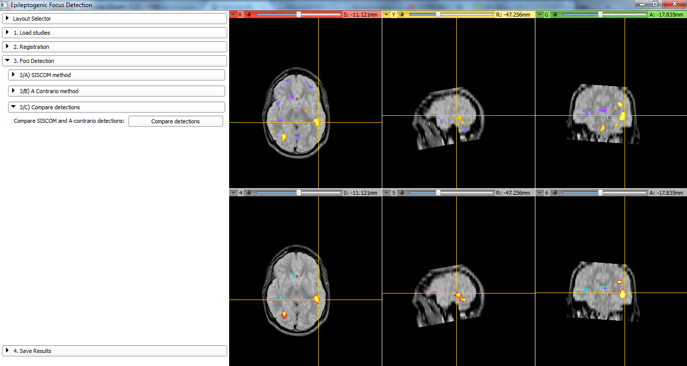
Compare candidates obtained using SISCOM and a-contrario method.
Tutorials
N/A
Panels and their use
- Load Studies: The following studies are required:
- SPECT basal
- SPECT ictal
- MRI (optional: for visualization of the results or SPECT/MRI registration)
- Registration: To compare the basal SPECT and the icatl SPECT the volumes must be registered. In this step it is possible to perform:
- SPECT basal/SPECT ictal registration
- SPECT/MRI registration (if MRI Available)
- SPECT basal/SPECT ictal mask (intersection) computation
- Foci detection: Differential (SISCOM) and a-contrario methods are implemented. Results can be visualized in MRI Space (if available) or SPECT Space. Results can be compared.
- Differential method: Given two SPECT images (basal and ictal) in the same space (already registered), the images are substracted. An intensity normalization step is applied.
- A-contrario: to be described
Similar Modules
N/A
References
N/A
Information for Developers
| Section under construction. |
