Difference between revisions of "Documentation/4.3/Announcements"
| Line 62: | Line 62: | ||
<gallery caption="New and Improved Extensions" widths="250px" heights="150px" perrow="4"> | <gallery caption="New and Improved Extensions" widths="250px" heights="150px" perrow="4"> | ||
| + | Image:AirwaySegmentation Module.png|[[Documentation/{{documentation/version}}/Extensions/AirwaySegmentation|Airway Segmentation]] {{new}} | ||
| + | Image:CMRTK-logo.png|[[Documentation/{{documentation/version}}/Extensions/CARMA|Cardiac MRI Toolkit]] to analyze cardiac LGE-MRI images. {{new}} | ||
| + | Image:ChangeTracker logo.png|[[Documentation/{{documentation/version}}/Extensions/ChangeTracker| Change Tracker]] for quantification of the subtle changes in pathology. {{new}} | ||
| + | Image:DiceSimilarityCoefficient.png|[[Documentation/{{documentation/version}}/Extensions/DiceComputation|Dice Computation]] is capable of calculating the Dice Similarity Coefficient (DSC) between multiple label map images. {{new}} | ||
| + | Image:ErodeDilateLabelIcon.png|[[Documentation/{{documentation/version}}/Extensions/ErodeDilateLabel|Erode/Dilate Label]] to perform binary erosion and dilation of a specified label. {{new}} | ||
| + | Image:IASEM-Screenshot1.png|[[Documentation/{{documentation/version}}/Extensions/IASEM| IASEM]] for segmentation and processing of IASEM Electron Microscopy images. {{new}} | ||
Image:QuickToolsLogo.png|[[Documentation/{{documentation/version}}/Extensions/ImageMaker|Image Maker]] to create volumes from scratch. {{new}} | Image:QuickToolsLogo.png|[[Documentation/{{documentation/version}}/Extensions/ImageMaker|Image Maker]] to create volumes from scratch. {{new}} | ||
| + | Image:LAScarSegmenter.png|[[Documentation/{{documentation/version}}/Extensions/LAScarSegmenter|LA Scar Segmenter]] to perform segmentation of the left atrial scarring tissue from MR images. {{new}} | ||
| + | Image:LASegmenter.png|[[Documentation/{{documentation/version}}/Extensions/LASegmenter|LA Segmenter]] to perform segmentation of the left atrium from MR images. {{new}} | ||
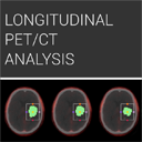
| + | Image:LongitudinalPETCTLogo.png|[[Documentation/{{documentation/version}}/Extensions/LongitudinalPETCT| Longitudinal PET/CT]] to provide a user friendly Slicer interface for quantification of DICOM PET/CT image data {{updated} | ||
| + | Image:MatlabBridgeLogo.png|[[Documentation/{{documentation/version}}/Extensions/MatlabBridge| Matlab Bridge]] to allow running Matlab functions directly in 3D Slicer. {{new}} | ||
| + | Image:ModelClipScreenShot.png|[[Documentation/{{documentation/version}}/Extensions/ModelClip| Model Clip]] to set the osteotomy trajectory with multiple planes, and clip the model with just one click. {{new}} | ||
| + | Image:PathXplorer PathCreation.png|[[Documentation/{{documentation/version}}/Extensions/PathPlanner| Path Planner]] designed to facilitate the creation of trajectory. {{new}} | ||
| + | Image:PlusRemoteLogo.png|[[Documentation/{{documentation/version}}/Extensions/PlusRemote|PlusRemote]] for sending commands through OpenIGTLink Remote to PLUS. {{new}} | ||
| + | Image:Portplacement.png|[[Documentation/{{documentation/version}}/Extensions/PortPlacement| Port Placement]] assists in the planning of surgical port placement in a laparoscopic procedure. {{new}} | ||
| + | Image:ReportingLogo.png|[[Documentation/{{documentation/version}}/Extensions/Reporting|Reporting]] to create image annotations/markup that are stored in a structured form. {{updated}} | ||
| + | Image:SobolevSegmenter.png|[[Documentation/{{documentation/version}}/Extensions/SobolevSegmenter|Sobolev Segmenter]] implements Sobolev inner product based active contour, using the Chan-Vese energy functional. {{updated}} | ||
| + | Image:SlicerIGTLogo.png|[[Documentation/{{documentation/version}}/Extensions/SlicerIGT|SlicerIGT]] to use all the advanced features of 3D Slicer for real-time navigation. {{new}} | ||
| + | Image:SlicerRT Logo 2.0 128x128.png|[[Documentation/{{documentation/version}}/Extensions/SlicerRT|SlicerRT]] a tool for powerful radiotherapy research. {{updated}} | ||
Image:SlicerToKiwiExporterLogo.png|[[Documentation/{{documentation/version}}/Extensions/SlicerToKiwiExporter|SlicerToKiwiExporter]] provides users with any easy way to export [[Documentation/{{documentation/version}}/Modules/Models|models]] into a [http://www.kiwiviewer.org/ KiwiViewer]. {{new}} | Image:SlicerToKiwiExporterLogo.png|[[Documentation/{{documentation/version}}/Extensions/SlicerToKiwiExporter|SlicerToKiwiExporter]] provides users with any easy way to export [[Documentation/{{documentation/version}}/Modules/Models|models]] into a [http://www.kiwiviewer.org/ KiwiViewer]. {{new}} | ||
| − | Image: | + | Image:SwissSkullStripper.png|[[Documentation/{{documentation/version}}/Extensions/SwissSkullStripper|Swiss Skull Stripper]] registers an atlas image to the patient data. {{new}} |
| − | + | Image:TrackerStabilizer.png|[[Documentation/{{documentation/version}}/Extensions/TrackerStabilizer|Tracker Stabilizer]] allows to output a filtered transform node based on an tracker input (transform node). {{new}} | |
| − | |||
| − | |||
| − | Image: | ||
| − | |||
Image:TransformVisualizerLogo.png|[[Documentation/{{documentation/version}}/Extensions/TransformVisualizer|Transform Visualizer]] to visualize any transform (linear transform, B-spline deformable transform, any other non-linear transform) or vector volume. {{new}} | Image:TransformVisualizerLogo.png|[[Documentation/{{documentation/version}}/Extensions/TransformVisualizer|Transform Visualizer]] to visualize any transform (linear transform, B-spline deformable transform, any other non-linear transform) or vector volume. {{new}} | ||
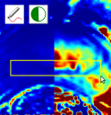
Image:WindowLevelEffectLogo.png|[[Documentation/{{documentation/version}}/Extensions/WindowLevelEffect|Window/Level Effect]] to adjust window/level for volumes using mouse and/or region of interest. {{new}} | Image:WindowLevelEffectLogo.png|[[Documentation/{{documentation/version}}/Extensions/WindowLevelEffect|Window/Level Effect]] to adjust window/level for volumes using mouse and/or region of interest. {{new}} | ||
| + | Image:XNATSlicer-Med.png|[[Documentation/{{documentation/version}}/Extensions/XNATSlicer| XNAT Slicer]] Secure GUI-based IO with any XNAT server. {{new}} | ||
Revision as of 17:13, 11 September 2013
Home < Documentation < 4.3 < Announcements
|
For the latest Slicer documentation, visit the read-the-docs. |
| Introduction | Our Philosophy | Slicer Highlights | Slicer Extensions | Other Improvements, Additions & Documentation |
Summary
The community of Slicer developers is proud to announce the release of Slicer 4.3.
- Slicer 4.3 introduces
- a new App store, called the extension manager, for adding capabilities to Slicer. Close to 40 plug-ins are currently available.
- close to 500 feature improvements and bug fixes lead to improved performance and stability.
- augmentation of many modules.
- improved workflows in registration and dMRI
- the multivolume explorer allows interaction with time series and DCE data.
- major improvements to the DICOM module
- a new Markups module to streamline the use of fiducial lists
- Click here to download Slicer 4.3 for different platforms and find pointers to the source code, mailing lists and bug tracker.
- Please note that Slicer continues to be a research package and is not intended for clinical use. Testing of functionality is an ongoing activity with high priority, however, some features of Slicer are not fully tested.
- The Slicer Training page provides a series of courses for learning how to use Slicer. The portfolio contains self-guided presentation and sample data sets.
The main slicer.org pages provide a guided tour to the application, training materials, and the development community. New users should start there because we try to keep the pages organized and up to date.
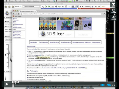
Find out more about Slicer 4.3 in the webinar held on [...]:
What is 3D Slicer
Slicer is a community platform created for the purpose of subject specific image analysis and visualization.
- Multi-modality imaging including, MRI, CT, US, nuclear medicine, and microscopy
- Multi organ from head to toe
- Bidirectional interface for devices
- Expandable and interfaced to multiple toolkits
There is no restriction on use, but permissions and compliance with rules are responsibility of users. For details on the license see here
Slicer 4.3 Highlights
- New and Improved Toolkits and Modules
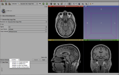
Added AutoRun mode for CLI modules to interactively search in parameter space and to create pipelines of filters. Watch the demo!
The Markups module replaces Annotation fiducials.
Slicer Extensions
- New and Improved Extensions
Cardiac MRI Toolkit to analyze cardiac LGE-MRI images. NEW
Change Tracker for quantification of the subtle changes in pathology. NEW
Dice Computation is capable of calculating the Dice Similarity Coefficient (DSC) between multiple label map images. NEW
Erode/Dilate Label to perform binary erosion and dilation of a specified label. NEW
IASEM for segmentation and processing of IASEM Electron Microscopy images. NEW
Image Maker to create volumes from scratch. NEW
LA Scar Segmenter to perform segmentation of the left atrial scarring tissue from MR images. NEW
LA Segmenter to perform segmentation of the left atrium from MR images. NEW
Longitudinal PET/CT to provide a user friendly Slicer interface for quantification of DICOM PET/CT image data {{updated}
Matlab Bridge to allow running Matlab functions directly in 3D Slicer. NEW
Model Clip to set the osteotomy trajectory with multiple planes, and clip the model with just one click. NEW
Path Planner designed to facilitate the creation of trajectory. NEW
PlusRemote for sending commands through OpenIGTLink Remote to PLUS. NEW
Port Placement assists in the planning of surgical port placement in a laparoscopic procedure. NEW
Reporting to create image annotations/markup that are stored in a structured form. UPDATED
Sobolev Segmenter implements Sobolev inner product based active contour, using the Chan-Vese energy functional. UPDATED
SlicerIGT to use all the advanced features of 3D Slicer for real-time navigation. NEW
SlicerRT a tool for powerful radiotherapy research. UPDATED
SlicerToKiwiExporter provides users with any easy way to export models into a KiwiViewer. NEW
Swiss Skull Stripper registers an atlas image to the patient data. NEW
Tracker Stabilizer allows to output a filtered transform node based on an tracker input (transform node). NEW
Transform Visualizer to visualize any transform (linear transform, B-spline deformable transform, any other non-linear transform) or vector volume. NEW
Window/Level Effect to adjust window/level for volumes using mouse and/or region of interest. NEW
XNAT Slicer Secure GUI-based IO with any XNAT server. NEW
Other Improvements, Additions & Documentation
Added advanced controls for the model display representation
Depth peeling option for translucent surface models in 3D views
Added read/write support for MRC files
For Developers: Looking at the Code Changes
From a git checkout you can easily see the all the commits since the time of the 4.4.2 release:
git log --since="Sat Dec 8 03:32:53 2012"
To see a summary of your own commits, you could use something like:
git log --since="Sat Dec 8 03:32:53 2012" --oneline --author=pieper
see the git log man page for more options.
Commit stats and full changelog
