Difference between revisions of "Documentation/Nightly/Modules/SegmentationAidedRegistration"
m (gallery resize) |
|||
| Line 48: | Line 48: | ||
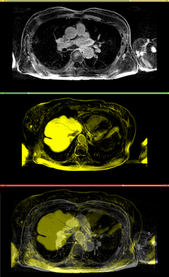
Image:AfibMIAffineRegistration.png|Affine registration using MI metric of the two LGE-MR images. Top: fixed image; Middle: registered moving image; Bottom: putting them together. | Image:AfibMIAffineRegistration.png|Affine registration using MI metric of the two LGE-MR images. Top: fixed image; Middle: registered moving image; Bottom: putting them together. | ||
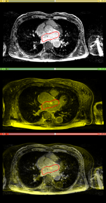
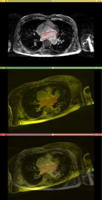
Image:AfibMIAffineRegistrationWithContour.png|Affine registration using MI metric of the two LGE-MR images, with the red contour showing the left atrium. Top: fixed image; Middle: registered moving image; Bottom: putting them together. We see that the overall registration is good, but the left atrium position is not accurate. In the case where we want to study the change of LA at those two time points, foucs has been put on the LA region. | Image:AfibMIAffineRegistrationWithContour.png|Affine registration using MI metric of the two LGE-MR images, with the red contour showing the left atrium. Top: fixed image; Middle: registered moving image; Bottom: putting them together. We see that the overall registration is good, but the left atrium position is not accurate. In the case where we want to study the change of LA at those two time points, foucs has been put on the LA region. | ||
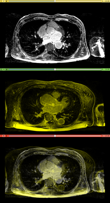
| + | Image:AfibSegmentationAidedRegistration.png|Segmentation aided registration using this module. The overall registration performance for the entire volume is bad, even worse than before registration. | ||
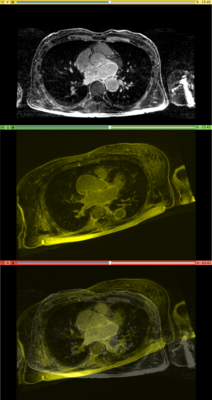
| + | Image:AfibSegmentationAidedRegistrationContour.png|Segmentation aided registration using this module. But at the target (left atrium) region, its very accurate, enabling analysis of the target region. | ||
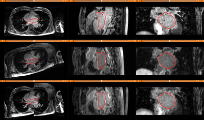
Image:SegmentationAidedRegistrationComparison.png|Results. Top row: Pre-op LGE-MRI with its segmentation of the left atrium (LA). Middle row: Pre-op LA contour overlayed on the registered Post-op LGE-MRI. See the matching of the LA region. Bottom row: checker-board view of Pre-op and registered Post-op LGE-MRIs: they match well at the LA region but badly otherwise, indicating the necessity of using segmentation to aid the registration. | Image:SegmentationAidedRegistrationComparison.png|Results. Top row: Pre-op LGE-MRI with its segmentation of the left atrium (LA). Middle row: Pre-op LA contour overlayed on the registered Post-op LGE-MRI. See the matching of the LA region. Bottom row: checker-board view of Pre-op and registered Post-op LGE-MRIs: they match well at the LA region but badly otherwise, indicating the necessity of using segmentation to aid the registration. | ||
</gallery> | </gallery> | ||
Revision as of 20:44, 18 June 2013
Home < Documentation < Nightly < Modules < SegmentationAidedRegistration
|
For the latest Slicer documentation, visit the read-the-docs. |
Introduction and Acknowledgements
|
This work is part of the National Alliance for Medical Image Computing (NA-MIC), funded by the National Institutes of Health through the NIH Roadmap for Medical Research, Grant U54 EB005149. Information on NA-MIC can be obtained from the NA-MIC website. Contributor: Yi Gao, Brigham and Women's Hospital | |||||||||
|
Module Description
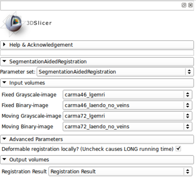
When we want to register two images, often times, we would like an accurate matching at certain region. For example, in the atrial fibrillation longitudinal study, we want to register the pre-op and post-op images. In particular, we want the matching at the atrium to be emphasized. In such cases, we can use the segmentation of the target region, atrium in the previous example, to aid the registration process. This module is such a segmentation aided registration tool.
Use Cases
- In the atrial fibrillation longitudinal study, we want to register the pre-op and post-op images. In particular, we want the matching at the atrium to be emphasized.
Affine registration using MI metric of the two LGE-MR images, with the red contour showing the left atrium. Top: fixed image; Middle: registered moving image; Bottom: putting them together. We see that the overall registration is good, but the left atrium position is not accurate. In the case where we want to study the change of LA at those two time points, foucs has been put on the LA region.
Results. Top row: Pre-op LGE-MRI with its segmentation of the left atrium (LA). Middle row: Pre-op LA contour overlayed on the registered Post-op LGE-MRI. See the matching of the LA region. Bottom row: checker-board view of Pre-op and registered Post-op LGE-MRIs: they match well at the LA region but badly otherwise, indicating the necessity of using segmentation to aid the registration.
Tutorials
N/A
Panels and their use
- Module panel
Similar Modules
N/A
References
- Y Gao, Y Rathi, S Bouix, A Tannenbaum; Filtering in the Diffeomorphism Group and the Registration of Point Sets; IEEE Transactions on Image Processing 21 (10), 4383--4396
- Y Gao, B Gholami, RS MacLeod, J Blauer, WM Haddad, A Tannenbaum; Segmentation of the Endocardial Wall of the Left Atrium using Local Region-Based Active Contours and Statistical Shape Learning. Proceedings of SPIE Medical Imaging 2010.
Information for Developers
| Section under construction. |


