Difference between revisions of "Stanford Simbios group"
From Slicer Wiki
| Line 15: | Line 15: | ||
==Atlas Generation from Input MR Images== | ==Atlas Generation from Input MR Images== | ||
===Model Generation from Input MR Images=== | ===Model Generation from Input MR Images=== | ||
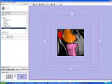
| − | Pre-Segmented models for femur, patella and tibia are obtained for a patient (in .stl format). This models can be prepared using the hypermesh software. The models contain vertices of triangles representing femur, tibia and patella regions. | + | Pre-Segmented models for femur, patella and tibia are obtained for a patient (in .stl format). This models can be prepared using the hypermesh software. The models contain vertices of triangles representing femur, tibia and patella regions. |
| + | [[Image:All_Three.JPG|left|thumb| Pre-Segmented Femur/Patella/Tibia Model]] | ||
| + | |||
===Create a filled label map using PolyDataToFilledLabelMap module in slicer=== | ===Create a filled label map using PolyDataToFilledLabelMap module in slicer=== | ||
===EM Segmentation based on the atlas=== | ===EM Segmentation based on the atlas=== | ||
Revision as of 18:03, 28 April 2009
Home < Stanford Simbios group
Contents
Aim
To develop a generic semi-automatic segmentation toolkit to convert images of musculoskeletal structures to 3D models.
Process Flowchart
Progress
Atlas Generation from Input MR Images
Model Generation from Input MR Images
Pre-Segmented models for femur, patella and tibia are obtained for a patient (in .stl format). This models can be prepared using the hypermesh software. The models contain vertices of triangles representing femur, tibia and patella regions.


