Difference between revisions of "Announcements:Slicer3.2"
| Line 22: | Line 22: | ||
=Highlights= | =Highlights= | ||
| − | <gallery caption="Slicer v3.2 - New and Improved Feature Highlights" widths="250px" heights="150px" perrow=" | + | <gallery caption="Slicer v3.2 - New and Improved Feature Highlights" widths="250px" heights="150px" perrow="3"> |
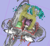
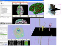
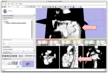
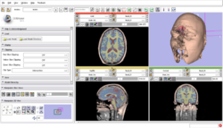
Image:ComplexVis.png|Complex Visualization Capabilities: Combining cross-sections and 3D surface models tumor, brain morphology, MR angiogram, fMRI and DTI) | Image:ComplexVis.png|Complex Visualization Capabilities: Combining cross-sections and 3D surface models tumor, brain morphology, MR angiogram, fMRI and DTI) | ||
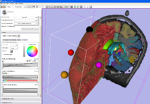
Image:VolRend.png|Volume Rendering: [[Modules:VolumeRendering-Documentation|Fully integrated volume rendering]] with cropping for easy exploration of volumetric data | Image:VolRend.png|Volume Rendering: [[Modules:VolumeRendering-Documentation|Fully integrated volume rendering]] with cropping for easy exploration of volumetric data | ||
| Line 30: | Line 30: | ||
Image:Editor-v3-2.png|[[Modules:Editor-Documentation|Interactive Editor:]] This new module allows interactive segmentation with robust 2D and 3D algorithms | Image:Editor-v3-2.png|[[Modules:Editor-Documentation|Interactive Editor:]] This new module allows interactive segmentation with robust 2D and 3D algorithms | ||
Image:IO.png|IO: [[Documentation|Improved IO capabilities]] include support for DICOM, NRRD, NIFTI, Tiff, JPG, Freesurfer, FITS and a number of other formats | Image:IO.png|IO: [[Documentation|Improved IO capabilities]] include support for DICOM, NRRD, NIFTI, Tiff, JPG, Freesurfer, FITS and a number of other formats | ||
| − | Image: | + | Image:DTI glyphs.png|Glyphs on corticospinal tract:<br>[[Slicer3:DTMRI|New Diffusion Imaging infrastructure]] includes, dicom import, gradient editor, visualiztion |
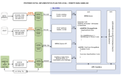
Image:DataLoadingStartPlan.png|[[Slicer3:Remote_Data_Handling|Remote Data Handling]] allows uploads and downloads from image informatics frameworks such as BIRN, and XNAT | Image:DataLoadingStartPlan.png|[[Slicer3:Remote_Data_Handling|Remote Data Handling]] allows uploads and downloads from image informatics frameworks such as BIRN, and XNAT | ||
Image:Slicer_IGTL_PartialImage.png|[http://wiki.na-mic.org/Wiki/index.php/IGT:ToolKit IGT Toolkit] to enable research in Image Guided Therapies | Image:Slicer_IGTL_PartialImage.png|[http://wiki.na-mic.org/Wiki/index.php/IGT:ToolKit IGT Toolkit] to enable research in Image Guided Therapies | ||
Revision as of 10:09, 16 June 2008
Home < Announcements:Slicer3.2Introduction
The community of Slicer developers is proud to announce the release of Slicer 3.2. This effort is the culmination of hundreds of person years and tens of millions of dollars of effort [1]. Slicer leverages state of the art open-source toolkits for visualization, medical image analysis, software process, and other tools for processing and accessing data (for more information see the description of the NA-MIC Kit). Slicer offers these capabilities as part of the open-source framework known as the NA-MIC Kit, which facilitates on-going research in biomedical computing, supports commercialization through NA-MIC Kit components, and provides a spectrum of capabilities suitable for researchers with varying levels of computer skills.
|
Slicer 3.2 is a general purpose biomedical computing application with extensive built-in visualization and analysis capabilities, accessible through an easy to use graphical interface. For advanced users, Slicer may be extended at run-time with user-defined plug-in modules. Release candidates for this application will be available the first week of June 2008. This new release contains hundreds of changes to the software. Highlights include:
Click here to download different versions of Slicer3 and find pointers to the source code, mailing lists and bug tracker. Please note that Slicer continues to be a research package and is not intended for clinical use. Testing of functionality is an ongoing activity with high priority, however, some features of Slicer3 are not fully tested. |
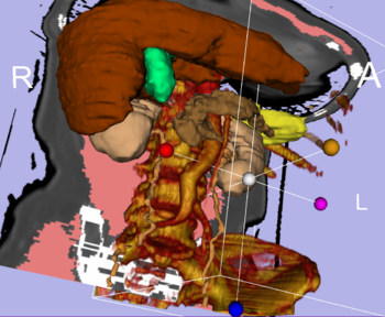
Integrated Volume Rendering: View of the abdominal atlas Bone and large vessels are volume rendered. |
Highlights
- Slicer v3.2 - New and Improved Feature Highlights
Volume Rendering: Fully integrated volume rendering with cropping for easy exploration of volumetric data
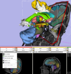
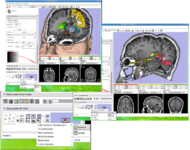
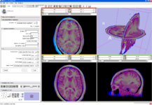
Implicit Slice Widget: An interactive tool for specifying oblique views (part of the VTK widget family).
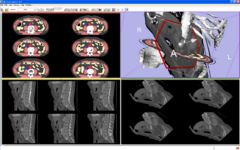
The Lightbox: Functionality for displaying cross-sectional data in columns and rows.
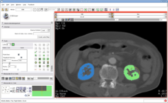
Interactive Editor: This new module allows interactive segmentation with robust 2D and 3D algorithms
IO: Improved IO capabilities include support for DICOM, NRRD, NIFTI, Tiff, JPG, Freesurfer, FITS and a number of other formats
Glyphs on corticospinal tract:
New Diffusion Imaging infrastructure includes, dicom import, gradient editor, visualiztionRemote Data Handling allows uploads and downloads from image informatics frameworks such as BIRN, and XNAT
IGT Toolkit to enable research in Image Guided Therapies
EM Segmenter: A configurable image segmentation tool that uses intensity distributions along with atlas information
Quality Software Process The NA-MIC Kit employs a test-driven software development process. Slicer3 also has a new build process.
Slicer in Numbers
The numbers in this table represent the components of the NA-MIC kit. Slicer 3 is based on the NA-MIC kit.
Source: http://www.ohloh.org
Captured on May 30 2008. The numbers in the column entitled "lines of code" are hard numbers. The other two columns are estimates based on some assumptions. Please see the Ohloh website for an explanation of how the numbers were computed.
| Package | Lines of code | Person years | Price tag at 100k per person year |
| Slicer | 587,919 | 161 | $16,068,440 |
| KWW | 189,627 | 49 | $ 4,925,590 |
| VTK | 1,344,989 | 385 | $38,521,873 |
| ITK | 711,474 | 197 | $19,712,495 |
| CMAKE | 213,671 | 56 | $ 5,586,895 |
| TEEM | 113,457 | 29 | $ 2,874,768 |
| Total | 3,161,137 | 877 | $87,690,061 |
