Difference between revisions of "Documentation/Nightly/Extensions/PercutaneousApproachAnalysis"
| Line 89: | Line 89: | ||
[3] Ido K, Isoda N, Sugano K. (2001) Microwave coagulation therapy for liver cancer: laparoscopic microwave coagulation. J Gastroenterol 36(3): 145-52<br> | [3] Ido K, Isoda N, Sugano K. (2001) Microwave coagulation therapy for liver cancer: laparoscopic microwave coagulation. J Gastroenterol 36(3): 145-52<br> | ||
[4] Khlebnikov R, Kainz B, Muehl J, Schmalstieg D. (2011) Crepscular Rays for Tumor Accessibility Planning. IEEE Trans Vis Comput Graph 17(12): 2163-72<br> | [4] Khlebnikov R, Kainz B, Muehl J, Schmalstieg D. (2011) Crepscular Rays for Tumor Accessibility Planning. IEEE Trans Vis Comput Graph 17(12): 2163-72<br> | ||
| + | [5] [http://www.slicer.org/slicerWiki/index.php/Documentation/Nightly/Extensions/PortPlacement PortPlacement module] | ||
| + | [6] [http://www.slicer.org/slicerWiki/index.php/Documentation/Nightly/Extensions/PortPlacement SPL Abdominal Atlas (used in examples)] | ||
<!-- ---------------------------- --> | <!-- ---------------------------- --> | ||
Revision as of 12:12, 1 December 2014
Home < Documentation < Nightly < Extensions < PercutaneousApproachAnalysis
|
For the latest Slicer documentation, visit the read-the-docs. |
Introduction and Acknowledgements
|
This work is supported by Shiga University of Medical Science (SUMS) in Japan, NA-MIC, NCIGT, and Slicer Community.
This work is also supported in part by the National Institute of Health (R01CA111288, P01CA067165, P41RR019703, P41EB015898, R01CA124377, R01CA138586, R42CA137886), and was presented in CARS 2014 meetingMedia:CARS2014 Murakami.pdf. | |||||||||
|
Module Description
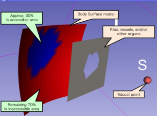
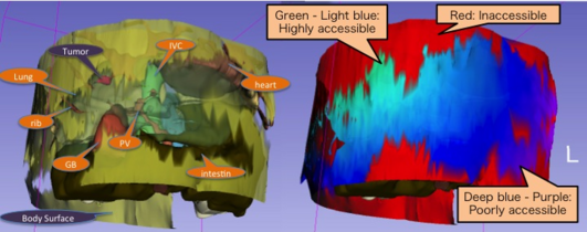
The Percutaneous Approach Analysis is used to calculate and visualize the accessibility of liver tumor with a percutaneous approach. The software uses the “ray-tracing” method in which the target tumor serves as a light source to identify the area of the skin that allows direct approach to the target tumor with a straight needle. This software takes the Body Surface model, Obstacle model, and a fiducial point in the liver model as inputs. The software also examines each line that connects the fiducial point and the center of each polygon cell on the Body Surface model and check if the line intersects the Obstacle model. If it does not intersect, the cell is identified as an accessible area. The polygon is colored based on the accessibility to visualize the results in 3D. For quantitative analysis, the software calculates the ratio of the total accessible area to the total area of the Body Surface model as the accessibility score (AS) for the specified fiducial point in the liver. Additionally, the software calculates and visualizes the distance between the target tumor and each polygon of the accessible area on the Body Surface model.
Use cases
- An example using a clinical model -
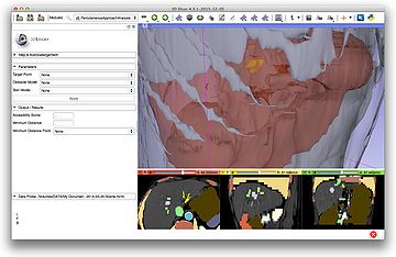
1. Model preparation
Preparing required 2 models as BodySurface and Obstacle

2. Put fiducial
Putting a fiducial point at target position
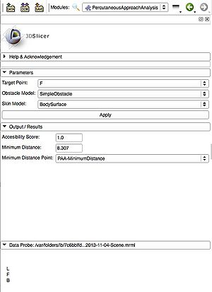
3. Input and Run
Selecting an appropriate object on each parameter pull-down window and push ‘Apply’ button
If you input ‘Minimum Distance Point’ in Output field, the nearest point from the target will be displayed on the Body Surface model.
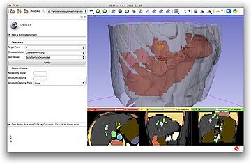
4. Output field
Accessibility score and Minimum Distance are displayed in the result field.

5. Scalar map
A color map showing approachable and inapproachable area is available.
To visualize it, a ‘visible’ check-box should be turned on in Scalar parameter of Models module.
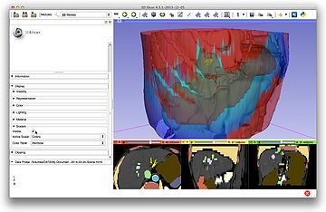
Tutorials
Typical use case is provided as tutorial slides.
Panels and Parameters
Parameters
- Target Point: Input an annotation fiducial point as a target of percutaneous approach.
- Obstacle Model: Input one Volume Model which you think as a barrier of needle insertion.
- Skin Model: Input one Volume Model on which the accessibile area should be measured as an inserting site.
Output/Results
- Accessibility Score: This box shows Accessibility Score which is calculated by accessibility area and total area on the selected Skin Model.
- Minimum Distance: The shortest distance between the Target Point and the surface of Skin Model.
- Minimum Distance Point: Input this when you want to know the nearest point on the Skin Model from the Target Point.
Similar Modules
N/A
References
[1] Rossi S, Di Stasi M, Buscarini E, Cavanna L, Quaretti P, Squassante E, Garbagnati F, Buscarini L. (1995) Percutaneous radiofrequency interstitial thermal ablation in the treatment of small hepatocellular carcinoma. Cancer J Sci Am 1(1): 73-81
[2] Silverman SG, Tuncali K, Adams DF, van Sonnenberg E, Zou KH, Kacher DF, Morrison PR, Jolesz FA. (2000) MR Imaging-guided Percutaneous Cryotherapy of Liver Tumors: Initial Experience. Radiology 217: 657-64
[3] Ido K, Isoda N, Sugano K. (2001) Microwave coagulation therapy for liver cancer: laparoscopic microwave coagulation. J Gastroenterol 36(3): 145-52
[4] Khlebnikov R, Kainz B, Muehl J, Schmalstieg D. (2011) Crepscular Rays for Tumor Accessibility Planning. IEEE Trans Vis Comput Graph 17(12): 2163-72
[5] PortPlacement module
[6] SPL Abdominal Atlas (used in examples)
Logo
|
Information for Developers
| Section under construction. |

