Difference between revisions of "Documentation/4.3/Training"
From Slicer Wiki
m |
|||
| Line 20: | Line 20: | ||
*Author: Sonia Pujol, Ph.D. | *Author: Sonia Pujol, Ph.D. | ||
*Audience: First time users who want a general introduction to the software. | *Audience: First time users who want a general introduction to the software. | ||
| + | *Based on: 3D Slicer version 4.0 | ||
|align="right"| | |align="right"| | ||
[[image:SlicerWelcome-image.png|250px|SlicerWelcome tutorial]] | [[image:SlicerWelcome-image.png|250px|SlicerWelcome tutorial]] | ||
| Line 30: | Line 31: | ||
*Author: Sonia Pujol, Ph.D. | *Author: Sonia Pujol, Ph.D. | ||
*Audience: First time users who want to discover Slicer in 4 minutes. | *Audience: First time users who want to discover Slicer in 4 minutes. | ||
| + | *Based on: 3D Slicer version 4.0 | ||
*The [[media:Slicer4minute.zip|Slicer4Minute dataset]] contains an MR scan of the brain and 3D reconstructions of the anatomy | *The [[media:Slicer4minute.zip|Slicer4Minute dataset]] contains an MR scan of the brain and 3D reconstructions of the anatomy | ||
|align="right"| | |align="right"| | ||
| Line 41: | Line 43: | ||
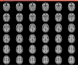
*Author: Sonia Pujol, Ph.D. | *Author: Sonia Pujol, Ph.D. | ||
*Audience: End-users | *Audience: End-users | ||
| + | *Based on: 3D Slicer version 4.1 | ||
*The [[Media:3DVisualizationData.zip | 3DVisualization dataset]] contain an MR scan and a series of 3D models of the brain. | *The [[Media:3DVisualizationData.zip | 3DVisualization dataset]] contain an MR scan and a series of 3D models of the brain. | ||
|align="right"| | |align="right"| | ||
| Line 53: | Line 56: | ||
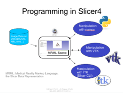
*The [[Media:Slicer4_ProgrammingTutorial_SPujol-SPieper.pdf | Hello Python Programming tutorial]] course guides through the integration of a python module in Slicer4. | *The [[Media:Slicer4_ProgrammingTutorial_SPujol-SPieper.pdf | Hello Python Programming tutorial]] course guides through the integration of a python module in Slicer4. | ||
*Author: Sonia Pujol, Ph.D., Steve Pieper, Ph.D. | *Author: Sonia Pujol, Ph.D., Steve Pieper, Ph.D. | ||
| − | *Audience: Developers | + | *Audience: Developers |
| + | *Based on: 3D Slicer version 4.1 | ||
*The [[Media:HelloPythonSlicer4.zip| HelloPython dataset]] contains three Python files and an MR scan of the brain. | *The [[Media:HelloPythonSlicer4.zip| HelloPython dataset]] contains three Python files and an MR scan of the brain. | ||
|align="right"| | |align="right"| | ||
| Line 67: | Line 71: | ||
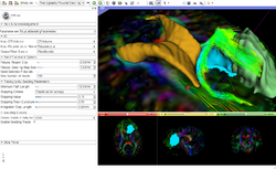
*Author: Sonia Pujol, Ph.D. | *Author: Sonia Pujol, Ph.D. | ||
*Audience: End-users and developers | *Audience: End-users and developers | ||
| + | *Based on: 3D Slicer version 4.1 | ||
*The [[Media:DiffusionMRI_tutorialData.zip |DTI dataset]] contains an MR Diffusion Weighted Imaging scan of the brain. | *The [[Media:DiffusionMRI_tutorialData.zip |DTI dataset]] contains an MR Diffusion Weighted Imaging scan of the brain. | ||
|align="right"| | |align="right"| | ||
| Line 77: | Line 82: | ||
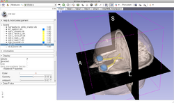
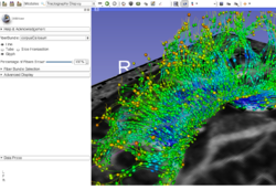
*The [[Media:NeurosurgicalPlanning_SoniaPujol.pdf | Neurosurgical Planning tutorial]] course guides through the generation of fiber tracts in the vicinity of a tumor. | *The [[Media:NeurosurgicalPlanning_SoniaPujol.pdf | Neurosurgical Planning tutorial]] course guides through the generation of fiber tracts in the vicinity of a tumor. | ||
*Author: Sonia Pujol, Ph.D., Ron Kikinis, M.D. | *Author: Sonia Pujol, Ph.D., Ron Kikinis, M.D. | ||
| − | *Audience: End-users and developers | + | *Audience: End-users and developers |
| + | *Based on: 3D Slicer version 4.1 | ||
*The [[Media:WhiteMatterExplorationData.zip| White Matter Exploration datasets]] contains a Diffusion Weighted Imaging scan of brain tumor patient. | *The [[Media:WhiteMatterExplorationData.zip| White Matter Exploration datasets]] contains a Diffusion Weighted Imaging scan of brain tumor patient. | ||
|align="right"| | |align="right"| | ||
| Line 88: | Line 94: | ||
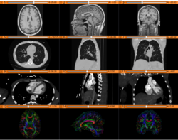
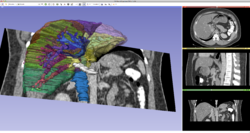
*The [[Media:3DVisualizationDICOMforRadiologyApplications_SoniaPujol_KittShaffer_RSNA2011.pdf | Slicer4RSNA]] course guides through 3D data loading and visualization of DICOM images for Radiology Applications in Slicer4. | *The [[Media:3DVisualizationDICOMforRadiologyApplications_SoniaPujol_KittShaffer_RSNA2011.pdf | Slicer4RSNA]] course guides through 3D data loading and visualization of DICOM images for Radiology Applications in Slicer4. | ||
*Author: Sonia Pujol, Ph.D., Kitt Shaffer, M.D., Ph.D. | *Author: Sonia Pujol, Ph.D., Kitt Shaffer, M.D., Ph.D. | ||
| − | *Audience: Radiologists and users of Slicer who need a more comprehensive overview over Slicer4 visualization capabilities. | + | *Audience: Radiologists and users of Slicer who need a more comprehensive overview over Slicer4 visualization capabilities. |
| + | *Based on: 3D Slicer version 4.0 | ||
*The [[Media:3DVisualizationData RSNA2011 part1.zip | Slicer4RSNAdataset1]] and [[Media:3DVisualizationData RSNA2011 part2.zip | Slicer4RSNAdataset2]] contain a series of MR and CT scans, and 3D models of the brain, lung and liver. | *The [[Media:3DVisualizationData RSNA2011 part1.zip | Slicer4RSNAdataset1]] and [[Media:3DVisualizationData RSNA2011 part2.zip | Slicer4RSNAdataset2]] contain a series of MR and CT scans, and 3D models of the brain, lung and liver. | ||
|align="right"| | |align="right"| | ||
[[Image:Slicer4RSNA_2.png|right|250px|]] | [[Image:Slicer4RSNA_2.png|right|250px|]] | ||
|} | |} | ||
| − | |||
==Slicer4 Quantitative Imaging tutorial== | ==Slicer4 Quantitative Imaging tutorial== | ||
| Line 100: | Line 106: | ||
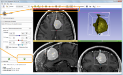
*The [[media:Slicer4QuantitativeImaging.pdf| Slicer4 Quantitative Imaging tutorial]] guides through the use for Slicer for quantifying small volumetric changes in slow-growing tumors, and for calculating Standardized Uptake Value (SUV) from PET/CT data. | *The [[media:Slicer4QuantitativeImaging.pdf| Slicer4 Quantitative Imaging tutorial]] guides through the use for Slicer for quantifying small volumetric changes in slow-growing tumors, and for calculating Standardized Uptake Value (SUV) from PET/CT data. | ||
*Authors: Jeffrey Yap, Ph.D., Ron Kikinis, M.D., Randy Gollub, M.D., Ph.D., Wendy Plesniak, Ph.D., Nicole Aucoin, B.Sc., Sonia Pujol, Ph.D., Valerie Humblet, Ph.D., Andriy Fedorov, Ph.D., Kilian Poh, Ph.D., Ender Konugolu, Ph.D. | *Authors: Jeffrey Yap, Ph.D., Ron Kikinis, M.D., Randy Gollub, M.D., Ph.D., Wendy Plesniak, Ph.D., Nicole Aucoin, B.Sc., Sonia Pujol, Ph.D., Valerie Humblet, Ph.D., Andriy Fedorov, Ph.D., Kilian Poh, Ph.D., Ender Konugolu, Ph.D. | ||
| − | *Audience: Radiologists and users of Slicer who need a more comprehensive overview over Slicer4 quantitative imaging capabilities. | + | *Audience: Radiologists and users of Slicer who need a more comprehensive overview over Slicer4 quantitative imaging capabilities. |
| + | *Based on: 3D Slicer version 4.0 | ||
*The [[media:PETCTFusion-TutorialData.zip| PETCTFusion]] and [[media:ChangeTracker2011.zip|Change Tracker]] datasets contain a series of MR, CT and PET data. | *The [[media:PETCTFusion-TutorialData.zip| PETCTFusion]] and [[media:ChangeTracker2011.zip|Change Tracker]] datasets contain a series of MR, CT and PET data. | ||
|align="right"| | |align="right"| | ||
[[Image:Slicer4_QuantitativeImaging.png|right|250px|]] | [[Image:Slicer4_QuantitativeImaging.png|right|250px|]] | ||
|} | |} | ||
| − | |||
=Summer 2012 Tutorial contest (under construction)= | =Summer 2012 Tutorial contest (under construction)= | ||
Revision as of 14:50, 6 September 2013
Home < Documentation < 4.3 < Training
|
For the latest Slicer documentation, visit the read-the-docs. |
Contents
Introduction: Slicer 4.3 Tutorials
- This page contains "How to" tutorials with matched sample data sets. They demonstrate how to use the 3D Slicer environment (version 4.3 release) to accomplish certain tasks.
- For tutorials for other versions of Slicer, please visit the Slicer training portal.
- For "reference manual" style documentation, please visit the Slicer 4.3 documentation page
- For questions related to the Slicer4 Compendium, please send an e-mail to Sonia Pujol, Ph.D
|
Some of these tutorials are based on older releases of 3D Slicer. The concepts are still useful but bear in mind that some interface elements and features will be different in updated versions. |
General Introduction
Slicer Welcome Tutorial
|
Slicer4Minute Tutorial
|
Slicer4 Data Loading and 3D Visualization
|
Tutorials for software developers
Slicer4 Programming Tutorial
|
Specific functions
Slicer4 Diffusion Tensor Imaging Tutorial
|
Slicer4 Neurosurgical Planning Tutorial
|
Slicer4 3D Visualization of DICOM images for Radiology Applications
|
Slicer4 Quantitative Imaging tutorial
|
Summer 2012 Tutorial contest (under construction)
Automatic Left Atrial Scar Segmenter
|
Qualitative and quantitative comparison of two RT dose distributions
|
Dose accumulation for adaptive radiation therapy
|
WebGL Export
|
OpenIGTLink
|
Additional resources
|
|
|
|
|
|
External Resources
Using the Editor
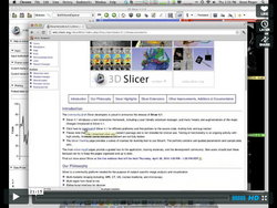
This set of tutorials about the use of slicer in paleontology is very well written and provides step-by-step instructions. Even though it covers slicer version 3.4, many of the concepts and techniques have applicability to the new version and to any 3D imaging field:
- Open Source Paleontologist: 3D Slicer: The Tutorial
- Open Source Paleontologist: 3D Slicer: The Tutorial Part II
- Open Source Paleontologist: 3D Slicer: The Tutorial Part III
- Open Source Paleontologist: 3D Slicer: The Tutorial Part IV
- Open Source Paleontologist: 3D Slicer: The Tutorial Part V
- Open Source Paleontologist: 3D Slicer: The Tutorial Part VI
Team Contributions
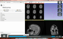
See the collection of videos on the Kitware vimeo album.
User Contributions
See the User Contributions Page for more content.
