Difference between revisions of "Documentation/4.0/Modules/IntensityDifferenceMetric"
From Slicer Wiki
(Created page with '<!-- ---------------------------- --> {{documentation/{{documentation/version}}/module-header}} <!-- ---------------------------- --> tumb|30px …') |
|||
| Line 34: | Line 34: | ||
* the images are aligned and the major source of difference is due to the anatomical changes of the structures | * the images are aligned and the major source of difference is due to the anatomical changes of the structures | ||
* the changes are small | * the changes are small | ||
| − | |[[Image:Slicer4_IntensityDifferenceMetric.png|thumb|450px| | + | * the structure of interest is hyperintensive in the baseline volume, and its segmentation is available |
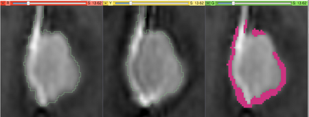
| + | |[[Image:Slicer4_IntensityDifferenceMetric.png|thumb|450px|Left to right: (1) baseline volume with the outline of the structure of interest; (2) followup volume co-registered with the baseline, with the same outline; (3) baseline volume with the voxels categorized as ''growth'' highlighted in red.]] | ||
|} | |} | ||
| Line 42: | Line 43: | ||
Most frequently used for these scenarios: | Most frequently used for these scenarios: | ||
* This metric is used in the last step of processing by [[Documentation/4.0/Modules/ChangeTracker|ChangeTracker]]. | * This metric is used in the last step of processing by [[Documentation/4.0/Modules/ChangeTracker|ChangeTracker]]. | ||
| + | * The metric can be used on its own, as long as the assumptions listed above are considered. | ||
<!-- ---------------------------- --> | <!-- ---------------------------- --> | ||
Revision as of 02:15, 24 November 2011
Home < Documentation < 4.0 < Modules < IntensityDifferenceMetric
Introduction and Acknowledgements
|
This work is supported by NA-MIC, NAC, NCIGT, and the Slicer Community. This work is partially supported by Brain Science Foundation and NIH U01 CA151261. | |||||||
|
Module Description
|
Intensity Difference Metric can be used to quantify the differences between the two images. The assumptions made by this metric are that:
|
Use Cases
Most frequently used for these scenarios:
- This metric is used in the last step of processing by ChangeTracker.
- The metric can be used on its own, as long as the assumptions listed above are considered.
Tutorials
See ChangeTracker documentation.
Panels and their use
Similar Modules
References
- Konukoglu, E., Wells, W. M., Novellas, S., Ayache, N., Kikinis, R., Black, P. M., & Pohl, K. M. (2008). Monitoring slowly evolving tumors. 2008 5th IEEE International Symposium on Biomedical Imaging: From Nano to Macro (pp. 812-815). IEEE. doi:10.1109/ISBI.2008.4541120 URL
- Pohl, K. M., Konukoglu, E., Novellas, S., Ayache, N., Fedorov, A., Talos, I.-F., Golby, A., et al. (2011). A new metric for detecting change in slowly evolving brain tumors: validation in meningioma patients. Neurosurgery, 68(1 Suppl Operative), 225-33. doi:10.1227/NEU.0b013e31820783d5 URL
Information for Developers
| Section under construction. |


