Difference between revisions of "Documentation/Nightly/Extensions/AnomalousFilters"
Acsenrafilho (talk | contribs) |
Acsenrafilho (talk | contribs) m Tag: 2017 source edit |
||
| (15 intermediate revisions by the same user not shown) | |||
| Line 8: | Line 8: | ||
{{documentation/{{documentation/version}}/module-introduction-start|{{documentation/modulename}} }} | {{documentation/{{documentation/version}}/module-introduction-start|{{documentation/modulename}} }} | ||
{{documentation/{{documentation/version}}/module-introduction-row}} | {{documentation/{{documentation/version}}/module-introduction-row}} | ||
| − | This work was partially funded by CAPES, a Brazillian | + | This work was partially funded by CAPES and CNPq, a Brazillian Agencies. Information on CAPES can be obtained on the [http://www.capes.gov.br/ CAPES website] and [http://www.cnpq.br/ CNPq website].<br> |
Author: Antonio Carlos da S. Senra Filho, CSIM Laboratory (University of Sao Paulo, Department of Computing and Mathematics)<br> | Author: Antonio Carlos da S. Senra Filho, CSIM Laboratory (University of Sao Paulo, Department of Computing and Mathematics)<br> | ||
Contact: Antonio Carlos da S. Senra Filho <email>acsenrafilho@usp.br</email><br> | Contact: Antonio Carlos da S. Senra Filho <email>acsenrafilho@usp.br</email><br> | ||
| Line 15: | Line 15: | ||
|Image:CSIM-logo.png|CSIM Laboratory | |Image:CSIM-logo.png|CSIM Laboratory | ||
|Image:USP-logo.png|University of Sao Paulo | |Image:USP-logo.png|University of Sao Paulo | ||
| + | |Image:CNPq-logo.png|CNPq Brazil | ||
|Image:CAPES-logo.png|CAPES Brazil | |Image:CAPES-logo.png|CAPES Brazil | ||
}} | }} | ||
| Line 21: | Line 22: | ||
<!-- ---------------------------- --> | <!-- ---------------------------- --> | ||
| − | {{documentation/{{documentation/version}}/module-section| | + | {{documentation/{{documentation/version}}/module-section|Extension Description}} |
{{documentation/{{documentation/version}}/module-description}} | {{documentation/{{documentation/version}}/module-description}} | ||
{| | {| | ||
[[Image:AnomalousDiffusionExtension-logo.png|left]] | [[Image:AnomalousDiffusionExtension-logo.png|left]] | ||
| − | Anomalous diffusion processes (ADP) are mathematically denoted by a power law in the Fokker-Planck equation, leading to the generalized form. There are several generalizations of the Fokker-Plank equation, which should give many different partial differential equations (PDEs). Here we | + | Anomalous diffusion processes (ADP) are mathematically denoted by a power law in the Fokker-Planck equation, leading to the generalized form. There are several generalizations of the Fokker-Plank equation, which should give many different partial differential equations (PDEs). Here we adopted the so-called porous media equation, allowing the super-diffusive and the sub-diffusive processes <ref>Tsallis, C. (2009). Introduction to Nonextensive Statistical Mechanics: Approaching a Complex World. Springer.</ref>. In porous media, channels are created promoting or blocking the flow of the density function, which has been proved to provide a suitable application for MRI noise attenuation <ref>Da S Senra Filho, A. C., Garrido Salmon, C. E., & Murta Junior, L. O. (2015). Anomalous diffusion process applied to magnetic resonance image enhancement. Physics in Medicine and Biology, 60(6), 2355–2373. doi:10.1088/0031-9155/60/6/2355</ref>. |
| − | Basically, there are two different filters already implementing the anomalous diffusion process: the isotropic anomalous diffusion and anisotropic anomalous diffusion filters | + | Basically, there are two different filters already implementing the anomalous diffusion process: the isotropic anomalous diffusion and anisotropic anomalous diffusion filters <ref>Da S Senra Filho, A. C., Garrido Salmon, C. E., & Murta Junior, L. O. (2015). Anomalous diffusion process applied to magnetic resonance image enhancement. Physics in Medicine and Biology, 60(6), 2355–2373. doi:10.1088/0031-9155/60/6/2355</ref>. These filters were already applied on different imaging MR modalities, such as structural T1 and T2 images <ref>Da S Senra Filho, A. C., Garrido Salmon, C. E., & Murta Junior, L. O. (2015). Anomalous diffusion process applied to magnetic resonance image enhancement. Physics in Medicine and Biology, 60(6), 2355–2373. doi:10.1088/0031-9155/60/6/2355</ref>, diffusion-weighted images (DWI and DTI)<ref>Senra Filho, A. C. da S., Duque, J. J., & Murta, L. O. (2013). Isotropic anomalous filtering in Diffusion-Weighted Magnetic Resonance Imaging. Conference Proceedings: Annual International Conference of the IEEE Engineering in Medicine and Biology Society. IEEE Engineering in Medicine and Biology Society. Conference, 2013, 4022–5. doi:10.1109/EMBC.2013.6610427</ref><ref>Senra Filho, A. C. da S., Simozo, F. H., Salmon, C. E. G., & Murta Junior, L. O. (2014). Anisotropic anomalous filter as a tool for decreasing patient exam time in diffusion-weighted MRI protocols. In XXIV Brazilian Congress on Biomedical Engineering (pp. 0–3). Uberlandia.</ref>, MRI relaxation T1 and T2 relaxometry<ref>Filho, A. C. da S. S., Barbosa, J. H. O., Salmon, C. E. G. S., & Junior, L. O. M. (2014). Anisotropic Anomalous Diffusion Filtering Applied to Relaxation Time Estimation in Magnetic Resonance Imaging. In Annual International Conference of the IEEE Engineering in Medicine and Biology Society (pp. 3893–3896). IEEE. doi:10.1109/EMBC.2014.6944474</ref> and to fMRI<ref>Filho, A. C. da S. S., Rondinoni, C., Santos, A. C. dos, & Junior, L. O. M. (2014). Brain Activation Inhomogeneity Highlighted by the Isotropic Anomalous Diffusion Filter. In Annual International Conference of the IEEE Engineering in Medicine and Biology Society (pp. 3313–3316). Chicago: IEEE. doi:10.1109/EMBC.2014.6944331</ref> as an initial study. |
<!-- ---------------------------- --> | <!-- ---------------------------- --> | ||
{{documentation/{{documentation/version}}/extension-section|Modules}} | {{documentation/{{documentation/version}}/extension-section|Modules}} | ||
| − | *[[Documentation/{{documentation/version}}/Modules/AADImageFilter|AAD Image Filter]] | + | * '''Structural image denoising with tissues border preservation function''': [[Documentation/{{documentation/version}}/Modules/AADImageFilter|AAD Image Filter]] |
| − | *[[Documentation/{{documentation/version}}/Modules/IADImageFilter|IAD Image Filter]] | + | * '''Structural image denoising without tissues border preservation function''': [[Documentation/{{documentation/version}}/Modules/IADImageFilter|IAD Image Filter]] |
| − | *[[Documentation/{{documentation/version}}/Modules/AADDiffusionWeightedData|AAD on DWI Image]] | + | * '''Diffusion-weighted MR image denoising with tissues border preservation''': [[Documentation/{{documentation/version}}/Modules/AADDiffusionWeightedData|AAD on DWI Image]] |
| + | * '''Echo-planar imaging denoising with tissues border preservation (fMRI and ASL)''': [[Documentation/{{documentation/version}}/Modules/AADEPIData|AAD on EPI Image]] | ||
<!-- ---------------------------- --> | <!-- ---------------------------- --> | ||
{{documentation/{{documentation/version}}/module-section|Use Cases}} | {{documentation/{{documentation/version}}/module-section|Use Cases}} | ||
Most frequently used for these scenarios: | Most frequently used for these scenarios: | ||
| − | * Use Case 1: Noise reduction as a | + | * Use Case 1: Noise reduction as a pre-processing step for tissue segmentation |
| − | **When dealing with single voxel classification schemes | + | **When dealing with single voxel classification schemes, a noise reduction pre-processing step is usually helpful to reduce data fluctuation due to acquisition artifacts (e.g. reducing the number of misclassified voxels). |
| − | * Use Case 2: | + | * Use Case 2: Volume rendering |
**Noise reduction will result in nicer looking volume renderings | **Noise reduction will result in nicer looking volume renderings | ||
* Use Case 3: Noise reduction as part of image processing pipeline | * Use Case 3: Noise reduction as part of image processing pipeline | ||
**Could offer a better segmentation and classification on specific brain image analysis such as in Multiple Sclerosis lesion segmentation | **Could offer a better segmentation and classification on specific brain image analysis such as in Multiple Sclerosis lesion segmentation | ||
| − | <gallery> | + | <gallery widths="400px" heights="400px" perrow="3"> |
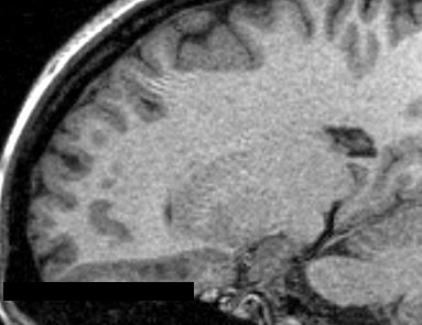
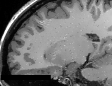
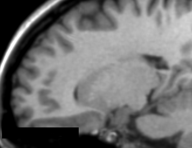
| + | Image:MRI_raw.png|Raw T1 weighted MRI Image | ||
Image:MRI_AAD.png|T1 weighted MRI Image with AAD filter (q=1.2) | Image:MRI_AAD.png|T1 weighted MRI Image with AAD filter (q=1.2) | ||
Image:MRI_IAD.png|T1 weighted MRI Image with IAD filter (q=1.2) | Image:MRI_IAD.png|T1 weighted MRI Image with IAD filter (q=1.2) | ||
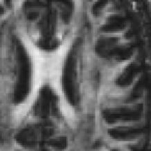
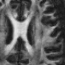
Image:DTI_FA_raw.png|DTI-FA map without image filtering process | Image:DTI_FA_raw.png|DTI-FA map without image filtering process | ||
Image:DTI_FA_AAD.png|DTI-FA map with AAD image filtering (q=0.4) | Image:DTI_FA_AAD.png|DTI-FA map with AAD image filtering (q=0.4) | ||
| − | |||
| − | |||
| − | |||
| − | |||
| − | |||
| − | |||
| − | |||
| − | |||
</gallery> | </gallery> | ||
<!-- ---------------------------- --> | <!-- ---------------------------- --> | ||
{{documentation/{{documentation/version}}/extension-section|Similar Extensions}} | {{documentation/{{documentation/version}}/extension-section|Similar Extensions}} | ||
| − | + | *[[Documentation/{{documentation/version}}/Modules/GradientAnisotropicDiffusion|Gradient Anisotropic Diffusion]] | |
<!-- ---------------------------- --> | <!-- ---------------------------- --> | ||
| Line 77: | Line 72: | ||
Repositories: | Repositories: | ||
| − | * Source code: [https://github.com/CSIM-Toolkits/ | + | * Source code: [https://github.com/CSIM-Toolkits/AnomalousFiltersExtension/ GitHub repository] |
| − | * Issue tracker: [https://github.com/CSIM-Toolkits/ | + | * Issue tracker: [https://github.com/CSIM-Toolkits/AnomalousFiltersExtension/issues open issues and enhancement requests] |
<!-- ---------------------------- --> | <!-- ---------------------------- --> | ||
{{documentation/{{documentation/version}}/extension-footer}} | {{documentation/{{documentation/version}}/extension-footer}} | ||
<!-- ---------------------------- --> | <!-- ---------------------------- --> | ||
Latest revision as of 15:45, 9 November 2024
Home < Documentation < Nightly < Extensions < AnomalousFilters
|
For the latest Slicer documentation, visit the read-the-docs. |
Introduction and Acknowledgements
|
This work was partially funded by CAPES and CNPq, a Brazillian Agencies. Information on CAPES can be obtained on the CAPES website and CNPq website. | |||||||||
|
Extension Description
Anomalous diffusion processes (ADP) are mathematically denoted by a power law in the Fokker-Planck equation, leading to the generalized form. There are several generalizations of the Fokker-Plank equation, which should give many different partial differential equations (PDEs). Here we adopted the so-called porous media equation, allowing the super-diffusive and the sub-diffusive processes [1]. In porous media, channels are created promoting or blocking the flow of the density function, which has been proved to provide a suitable application for MRI noise attenuation [2].
Basically, there are two different filters already implementing the anomalous diffusion process: the isotropic anomalous diffusion and anisotropic anomalous diffusion filters [3]. These filters were already applied on different imaging MR modalities, such as structural T1 and T2 images [4], diffusion-weighted images (DWI and DTI)[5][6], MRI relaxation T1 and T2 relaxometry[7] and to fMRI[8] as an initial study.
Modules
- Structural image denoising with tissues border preservation function: AAD Image Filter
- Structural image denoising without tissues border preservation function: IAD Image Filter
- Diffusion-weighted MR image denoising with tissues border preservation: AAD on DWI Image
- Echo-planar imaging denoising with tissues border preservation (fMRI and ASL): AAD on EPI Image
Use Cases
Most frequently used for these scenarios:
- Use Case 1: Noise reduction as a pre-processing step for tissue segmentation
- When dealing with single voxel classification schemes, a noise reduction pre-processing step is usually helpful to reduce data fluctuation due to acquisition artifacts (e.g. reducing the number of misclassified voxels).
- Use Case 2: Volume rendering
- Noise reduction will result in nicer looking volume renderings
- Use Case 3: Noise reduction as part of image processing pipeline
- Could offer a better segmentation and classification on specific brain image analysis such as in Multiple Sclerosis lesion segmentation
Similar Extensions
References
- da S Senra Filho, A.C., Garrido Salmon, C.E. & Murta Junior, L.O., 2015. Anomalous diffusion process applied to magnetic resonance image enhancement. Physics in Medicine and Biology, 60(6), pp.2355–2373. DOI: 10.1088/0031-9155/60/6/2355
- Filho, A.C. da S.S. et al., 2014. Anisotropic Anomalous Diffusion Filtering Applied to Relaxation Time Estimation in Magnetic Resonance Imaging. In Annual International Conference of the IEEE Engineering in Medicine and Biology Society. IEEE, pp. 3893–3896.
- Filho, A.C. da S.S., Barizon, G.C. & Junior, L.O.M., 2014. Myocardium Segmentation Improvement with Anisotropic Anomalous Diffusion Filter Applied to Cardiac Magnetic Resonance Imaging. In Annual Meeting of Computing in Cardiology.
- Filho, A.C. da S.S. et al., 2014. Brain Activation Inhomogeneity Highlighted by the Isotropic Anomalous Diffusion Filter. In Annual International Conference of the IEEE Engineering in Medicine and Biology Society. Chicago: IEEE, pp. 3313–3316.
- Senra Filho, A.C. da S., Duque, J.J. & Murta, L.O., 2013. Isotropic anomalous filtering in Diffusion-Weighted Magnetic Resonance Imaging. I. E. in M. and B. Society, ed. Conference proceedings : ... Annual International Conference of the IEEE Engineering in Medicine and Biology Society. IEEE Engineering in Medicine and Biology Society. Conference, 2013, pp.4022–5.
Information for Developers
| Section under construction. |
Repositories:
- Source code: GitHub repository
- Issue tracker: open issues and enhancement requests
- ↑ Tsallis, C. (2009). Introduction to Nonextensive Statistical Mechanics: Approaching a Complex World. Springer.
- ↑ Da S Senra Filho, A. C., Garrido Salmon, C. E., & Murta Junior, L. O. (2015). Anomalous diffusion process applied to magnetic resonance image enhancement. Physics in Medicine and Biology, 60(6), 2355–2373. doi:10.1088/0031-9155/60/6/2355
- ↑ Da S Senra Filho, A. C., Garrido Salmon, C. E., & Murta Junior, L. O. (2015). Anomalous diffusion process applied to magnetic resonance image enhancement. Physics in Medicine and Biology, 60(6), 2355–2373. doi:10.1088/0031-9155/60/6/2355
- ↑ Da S Senra Filho, A. C., Garrido Salmon, C. E., & Murta Junior, L. O. (2015). Anomalous diffusion process applied to magnetic resonance image enhancement. Physics in Medicine and Biology, 60(6), 2355–2373. doi:10.1088/0031-9155/60/6/2355
- ↑ Senra Filho, A. C. da S., Duque, J. J., & Murta, L. O. (2013). Isotropic anomalous filtering in Diffusion-Weighted Magnetic Resonance Imaging. Conference Proceedings: Annual International Conference of the IEEE Engineering in Medicine and Biology Society. IEEE Engineering in Medicine and Biology Society. Conference, 2013, 4022–5. doi:10.1109/EMBC.2013.6610427
- ↑ Senra Filho, A. C. da S., Simozo, F. H., Salmon, C. E. G., & Murta Junior, L. O. (2014). Anisotropic anomalous filter as a tool for decreasing patient exam time in diffusion-weighted MRI protocols. In XXIV Brazilian Congress on Biomedical Engineering (pp. 0–3). Uberlandia.
- ↑ Filho, A. C. da S. S., Barbosa, J. H. O., Salmon, C. E. G. S., & Junior, L. O. M. (2014). Anisotropic Anomalous Diffusion Filtering Applied to Relaxation Time Estimation in Magnetic Resonance Imaging. In Annual International Conference of the IEEE Engineering in Medicine and Biology Society (pp. 3893–3896). IEEE. doi:10.1109/EMBC.2014.6944474
- ↑ Filho, A. C. da S. S., Rondinoni, C., Santos, A. C. dos, & Junior, L. O. M. (2014). Brain Activation Inhomogeneity Highlighted by the Isotropic Anomalous Diffusion Filter. In Annual International Conference of the IEEE Engineering in Medicine and Biology Society (pp. 3313–3316). Chicago: IEEE. doi:10.1109/EMBC.2014.6944331


