Difference between revisions of "Documentation/Nightly/Extensions/OpenCAD"
From Slicer Wiki
| (10 intermediate revisions by 2 users not shown) | |||
| Line 21: | Line 21: | ||
{{documentation/{{documentation/version}}/module-introduction-end}} | {{documentation/{{documentation/version}}/module-introduction-end}} | ||
| + | |||
<!-- ---------------------------- --> | <!-- ---------------------------- --> | ||
| − | {{documentation/{{documentation/version}}/ | + | {{documentation/{{documentation/version}}/extension-section|Modules}} |
| − | + | *[[Documentation/{{documentation/version}}/Modules/SegmentCAD|SegmentCAD: Tumor Segmentation from DCE-MRI]] | |
| + | *[[Documentation/{{documentation/version}}/Modules/HeterogeneityCAD|HeterogeneityCAD: Feature Extraction toolbox for image heterogeneity analysis]] | ||
| − | <!-- ---------------------------- --> | + | <!-- ---------------------------- --> |
| − | {{documentation/{{documentation/version}}/ | + | {{documentation/{{documentation/version}}/extension-section|Extension Description}} |
| − | + | *The SegmentCAD module is designed to segment tumors from DCE-MRI datasets which include a pre-contrast image and post-contrast images at different time points. | |
| − | + | **SegmentCAD uses blackbox methods to calculate the wash-in and wash-out slopes from the time-intensity curves. | |
| − | * | + | **The segmentation output is a Label Map with red, yellow, and blue colors respectively identifying washout (Type III), plateau (Type II), and persistent (Type I) voxels. |
| − | * | + | *The HeterogeneityCAD module is an extensible, image feature extraction toolbox primarily to quantify the heterogeneity of tumor images and their label maps. |
| − | * | + | **Metrics have been implemented from a variety of feature classes including: |
| − | ** | + | ***First-Order/Histogram statistics |
| − | *** | + | ***Morphology/Shape measures and Geometrical (4D Extrusion) measures |
| − | ** | + | ***Renyi/Fractal dimensions |
| − | * | + | ***Texture features computed from Gray-Level Co-occurrence Matrices (GLCM) and from Gray-Level Run Length matrices (GLRL) |
| − | ** | + | |
| − | *** | ||
| − | |||
| − | |||
| − | <!-- ---------------------------- | + | <!-- ---------------------------- |
{{documentation/{{documentation/version}}/module-section|Features}} | {{documentation/{{documentation/version}}/module-section|Features}} | ||
| − | + | --> | |
| − | |||
| − | |||
| − | |||
<!-- ---------------------------- --> | <!-- ---------------------------- --> | ||
{{documentation/{{documentation/version}}/module-section|Tutorials}} | {{documentation/{{documentation/version}}/module-section|Tutorials}} | ||
| − | [[Media: | + | *SegmentCAD |
| + | **[[Media:SegmentCADTutorial.pptx|SegmentCAD Tutorial (pptx)]] | ||
<!-- ---------------------------- --> | <!-- ---------------------------- --> | ||
{{documentation/{{documentation/version}}/module-section|Data sets}} | {{documentation/{{documentation/version}}/module-section|Data sets}} | ||
| − | [[Media:Breast-data1.zip|Breast DCE-MRI Data Set 1 (zip file containing the nrrd volumes for the | + | *SegmentCAD |
| − | + | **[[Media:Breast-data1.zip|Breast DCE-MRI Data Set 1 (zip file containing the nrrd volumes for the tutorial)]] | |
| − | [[Media:Breast-data2.zip|Breast DCE-MRI Data Set 2 (zip file containing additional test set of nrrd volumes)]] | + | **[[Media:Breast-data2.zip|Breast DCE-MRI Data Set 2 (zip file containing additional test set of nrrd volumes)]] |
| − | + | *HeterogeneityCAD | |
| − | + | **[[Media:BreastHeteroCADData.zip|Breast DCE-MRI Data Set (zip file containing the nrrd volumes for the tutorial)]] | |
| − | |||
| − | |||
| − | |||
| − | |||
| − | |||
| − | * | ||
| − | ** | ||
| − | |||
| − | |||
| − | |||
| − | |||
| − | |||
| − | |||
| − | |||
| − | |||
| − | |||
| − | |||
| − | |||
| − | |||
| − | |||
| − | |||
| − | |||
| − | |||
| − | |||
| − | |||
| − | |||
| − | |||
| − | |||
| − | |||
| − | |||
| − | |||
| − | |||
<!-- ---------------------------- --> | <!-- ---------------------------- --> | ||
{{documentation/{{documentation/version}}/module-section|Quick Instructions for Use}} | {{documentation/{{documentation/version}}/module-section|Quick Instructions for Use}} | ||
| − | *Select the pre-contrast volume | + | *[[Documentation/{{documentation/version}}/Modules/SegmentCAD|SegmentCAD (Click link for detailed description)]] |
| − | *Select the first post-contrast volume | + | **Select the pre-contrast volume |
| − | *Select the second post-contrast volume | + | **Select the first post-contrast volume |
| − | *Select the third post-contrast volume | + | **Select the second post-contrast volume |
| − | *Select the fourth post-contrast volume | + | **Select the third post-contrast volume |
| − | + | **Select the fourth post-contrast volume | |
| − | *Create or select a label map volume node to represent the output of the segmentation | + | **Create or select a label map volume node to represent the output of the segmentation |
| − | + | **Click "Apply OpenCAD Segmentation" | |
| − | *Click "Apply OpenCAD Segmentation" | + | *[[Documentation/{{documentation/version}}/Modules/HeterogeneityCAD|HeterogeneityCAD (Click link for detailed description)]] |
| − | + | **Add an image or parameter map (.nrrd file) to the Nodes List | |
| − | + | **Select a corresponding segmentation label map to use as ROI | |
| − | + | **Click "Apply HeterogeneityCAD" | |
| − | |||
<!-- ---------------------------- --> | <!-- ---------------------------- --> | ||
{{documentation/{{documentation/version}}/module-section|Similar Modules}} | {{documentation/{{documentation/version}}/module-section|Similar Modules}} | ||
| − | + | *SegmentCAD: | |
| + | *HeterogeneityCAD: | ||
| + | **LabelStatistics | ||
<!-- ---------------------------- --> | <!-- ---------------------------- --> | ||
{{documentation/{{documentation/version}}/module-section|References}} | {{documentation/{{documentation/version}}/module-section|References}} | ||
| − | N | + | * J. Jayender, E. Gombos, S. Chikarmane, D. Dabydeen, F. A. Jolesz, and K. G. Vosburgh, “Statistical Learning Algorithm for In-situ and Invasive Breast Carcinoma Segmentation”, Journal of Computerized Medical Imaging and Graphics, vol. 37, no. 4, pp. 281-292, 2013 |
| + | * J. Jayender, S. A. Chikarmane, F. A. Jolesz and E. Gombos, “Automatic Segmentation of Invasive Breast Carcinomas from DCE-MRI using Time Series Analysis”, Journal of MRI, Article first published online 23 September 2013, doi: 10.1002/jmri.24394 | ||
| + | * J. Jayender, K.G. Vosburgh, E. Gombos, A. Ashraf, D. Kontos, S.C. Gavenonis, F. A. Jolesz and K. Pohl , “Automatic Segmentation of Breast Carcinomas from DCE-MRI using a Statistical Learning Algorithm”, IEEE International Symposium on Biomedical Imaging, pp. 122-125, 2012. | ||
| + | * J. Jayender, D.T. Ruan, V. Narayan, N. Agrawal, F. A. Jolesz and H. Mamata, “Segmentation of Parathyroid Tumors from DCE-MRI using Linear Dynamic System Analysis”, IEEE International Symposium on Biomedical Imaging, 2013. | ||
| + | * J. Jayender, J. Jagannathan, S.Chikarmane, C.P.Raut and F.A. Jolesz, “Computer-Aided Diagnosis of Breast Angiosarcoma: Results in 14 cases”, Quantitative Medical Imaging Symposium, 2013 (invited paper). | ||
| + | * HJWL Aerts, ER Velazquez, RTH Leijenaar, et al., "Decoding tumour phenotype by noninvasive imaging using a quantitative radiomics approach", vol. 5, Nat Communication, 2014. | ||
| + | |||
<!-- ---------------------------- --> | <!-- ---------------------------- --> | ||
{{documentation/{{documentation/version}}/module-section|Information for Developers}} | {{documentation/{{documentation/version}}/module-section|Information for Developers}} | ||
| − | |||
| − | |||
Source code: https://github.com/vnarayan13/Slicer-OpenCAD | Source code: https://github.com/vnarayan13/Slicer-OpenCAD | ||
Latest revision as of 03:46, 2 August 2014
Home < Documentation < Nightly < Extensions < OpenCAD
|
For the latest Slicer documentation, visit the read-the-docs. |
Introduction and Acknowledgements
|
This work is supported by NA-MIC, NCIGT, and the Slicer Community. | |||||||
This project is supported by P41 RR019703/RR/NCRR NIH HHS/United States, P01 CA067165/CA/NCI NIH HHS/United States and P41 EB015898/EB/NIBIB NIH HHS/United States |
Modules
- SegmentCAD: Tumor Segmentation from DCE-MRI
- HeterogeneityCAD: Feature Extraction toolbox for image heterogeneity analysis
Extension Description
- The SegmentCAD module is designed to segment tumors from DCE-MRI datasets which include a pre-contrast image and post-contrast images at different time points.
- SegmentCAD uses blackbox methods to calculate the wash-in and wash-out slopes from the time-intensity curves.
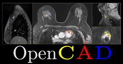
- The segmentation output is a Label Map with red, yellow, and blue colors respectively identifying washout (Type III), plateau (Type II), and persistent (Type I) voxels.
- The HeterogeneityCAD module is an extensible, image feature extraction toolbox primarily to quantify the heterogeneity of tumor images and their label maps.
- Metrics have been implemented from a variety of feature classes including:
- First-Order/Histogram statistics
- Morphology/Shape measures and Geometrical (4D Extrusion) measures
- Renyi/Fractal dimensions
- Texture features computed from Gray-Level Co-occurrence Matrices (GLCM) and from Gray-Level Run Length matrices (GLRL)
- Metrics have been implemented from a variety of feature classes including:
Tutorials
- SegmentCAD
Data sets
- SegmentCAD
- HeterogeneityCAD
Quick Instructions for Use
- SegmentCAD (Click link for detailed description)
- Select the pre-contrast volume
- Select the first post-contrast volume
- Select the second post-contrast volume
- Select the third post-contrast volume
- Select the fourth post-contrast volume
- Create or select a label map volume node to represent the output of the segmentation
- Click "Apply OpenCAD Segmentation"
- HeterogeneityCAD (Click link for detailed description)
- Add an image or parameter map (.nrrd file) to the Nodes List
- Select a corresponding segmentation label map to use as ROI
- Click "Apply HeterogeneityCAD"
Similar Modules
- SegmentCAD:
- HeterogeneityCAD:
- LabelStatistics
References
- J. Jayender, E. Gombos, S. Chikarmane, D. Dabydeen, F. A. Jolesz, and K. G. Vosburgh, “Statistical Learning Algorithm for In-situ and Invasive Breast Carcinoma Segmentation”, Journal of Computerized Medical Imaging and Graphics, vol. 37, no. 4, pp. 281-292, 2013
- J. Jayender, S. A. Chikarmane, F. A. Jolesz and E. Gombos, “Automatic Segmentation of Invasive Breast Carcinomas from DCE-MRI using Time Series Analysis”, Journal of MRI, Article first published online 23 September 2013, doi: 10.1002/jmri.24394
- J. Jayender, K.G. Vosburgh, E. Gombos, A. Ashraf, D. Kontos, S.C. Gavenonis, F. A. Jolesz and K. Pohl , “Automatic Segmentation of Breast Carcinomas from DCE-MRI using a Statistical Learning Algorithm”, IEEE International Symposium on Biomedical Imaging, pp. 122-125, 2012.
- J. Jayender, D.T. Ruan, V. Narayan, N. Agrawal, F. A. Jolesz and H. Mamata, “Segmentation of Parathyroid Tumors from DCE-MRI using Linear Dynamic System Analysis”, IEEE International Symposium on Biomedical Imaging, 2013.
- J. Jayender, J. Jagannathan, S.Chikarmane, C.P.Raut and F.A. Jolesz, “Computer-Aided Diagnosis of Breast Angiosarcoma: Results in 14 cases”, Quantitative Medical Imaging Symposium, 2013 (invited paper).
- HJWL Aerts, ER Velazquez, RTH Leijenaar, et al., "Decoding tumour phenotype by noninvasive imaging using a quantitative radiomics approach", vol. 5, Nat Communication, 2014.
Information for Developers
Source code: https://github.com/vnarayan13/Slicer-OpenCAD


