Difference between revisions of "Documentation/4.2/Modules/PkModeling"
From Slicer Wiki
(Prepend documentation/versioncheck template. See http://na-mic.org/Mantis/view.php?id=2887) |
|||
| (One intermediate revision by one other user not shown) | |||
| Line 1: | Line 1: | ||
| + | <noinclude>{{documentation/versioncheck}}</noinclude> | ||
<!-- ---------------------------- --> | <!-- ---------------------------- --> | ||
{{documentation/{{documentation/version}}/module-header}} | {{documentation/{{documentation/version}}/module-header}} | ||
| Line 49: | Line 50: | ||
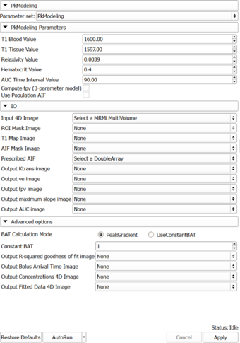
|[[Image:PkModelingUI061912.png|thumb|340px|PkModeling]] | |[[Image:PkModelingUI061912.png|thumb|340px|PkModeling]] | ||
| | | | ||
| − | * ''' | + | * '''IO''' |
** '''Input:''' 4D DCE-MRI data; 3D mask showing the location of the arterial input function. | ** '''Input:''' 4D DCE-MRI data; 3D mask showing the location of the arterial input function. | ||
** '''Output:''' 4 volumes showing the maps of quantitative parameters: ktrans, ve, maximum slope, and area under the curve (AUC). | ** '''Output:''' 4 volumes showing the maps of quantitative parameters: ktrans, ve, maximum slope, and area under the curve (AUC). | ||
Latest revision as of 07:45, 14 June 2013
Home < Documentation < 4.2 < Modules < PkModeling
|
For the latest Slicer documentation, visit the read-the-docs. |
Introduction and Acknowledgements
|
Extension: PkModeling | |||||
|
Module Description
|
PkModeling (Pharmacokinetics Modeling) calculates quantitative parameters from Dynamic Contrast Enhanced DCE-MRI images. This module performs two operations:
|
Use Cases
Tutorials
Panels and their use
|
Similar Modules
References
- Knopp MV, Giesel FL, Marcos H et al: Dynamic contrast-enhanced magnetic resonance imaging in oncology. Top Magn Reson Imaging, 2001; 12:301-308.
- Rijpkema M, Kaanders JHAM, Joosten FBM et al: Method for quantitative mapping of dynamic MRI contrast agent uptake in human tumors. J Magn Reson Imaging 2001; 14:457-463.
Information for Developers
| Section under construction. |


