Difference between revisions of "EMSegmenter-Tasks:MRI-Human-Brain-HIPR"
Belhachemi (talk | contribs) |
m (Text replacement - "\[http:\/\/www\.slicer\.org\/slicerWiki\/index\.php\/([^ ]+) ([^]]+)]" to "$2") |
||
| (2 intermediate revisions by one other user not shown) | |||
| Line 4: | Line 4: | ||
'''High In-Plane Resolution (HIPR)''' <br> | '''High In-Plane Resolution (HIPR)''' <br> | ||
Single channel automatic segmentation of T1 and T2 MRI brain scans into the major tissue classes (gray matter, white matter, csf). The task can be applied to T1 and T2 brain scan showing parts of the skull and neck. The pipeline consist of the following steps: | Single channel automatic segmentation of T1 and T2 MRI brain scans into the major tissue classes (gray matter, white matter, csf). The task can be applied to T1 and T2 brain scan showing parts of the skull and neck. The pipeline consist of the following steps: | ||
| − | * Step 1: Perform image inhomogeneity correction of the MRI scan via [ | + | * Step 1: Perform image inhomogeneity correction of the MRI scan via [[Modules:N4ITKBiasFieldCorrection-Documentation-3.6|N4ITKBiasFieldCorrection]] (Tustison et al 2010) |
* Step 2: Register the atlas to the MRI scan via [[Modules:BRAINSFit| BRAINSFit]] (Johnson et al 2007) | * Step 2: Register the atlas to the MRI scan via [[Modules:BRAINSFit| BRAINSFit]] (Johnson et al 2007) | ||
* Step 3: Compute the intensity distributions for each structure <BR> | * Step 3: Compute the intensity distributions for each structure <BR> | ||
| − | Compute intensity distribution (mean and variance) for each label by automatically sampling from the MR scan. The sampling for a specific label is constrained to the region that consists of voxels with high probability (top 95%) of being assigned to the label according to the aligned atlas. The alpha parameter for the EMSegmenter has been set to 0.01 to handle the high in-plane resolution of the scans. | + | Compute intensity distribution (mean and variance) for each label by automatically sampling from the MR scan. The sampling for a specific label is constrained to the region that consists of voxels with high probability (top 95%) of being assigned to the label according to the aligned atlas. '''The alpha parameter for the EMSegmenter has been set to 0.01 to handle the high in-plane resolution of the scans.''' |
* Step 4: Automatically segment the MRI scan into the structures of interest using [[Modules:EMSegmenter-3.6|EM Algorithm]] (Pohl et al 2007) | * Step 4: Automatically segment the MRI scan into the structures of interest using [[Modules:EMSegmenter-3.6|EM Algorithm]] (Pohl et al 2007) | ||
| Line 52: | Line 52: | ||
<gallery perrow=1: widths=1100px : heights=350px> | <gallery perrow=1: widths=1100px : heights=350px> | ||
| − | Image:MRI-Human-Brain-HIPR-T1Flair_map.png | + | Image:MRI-Human-Brain-HIPR-T1Flair_map.png|T1FLAIR |
| − | Image:MRI-Human-Brain-HIPR-T2Flair_map.png | + | Image:MRI-Human-Brain-HIPR-T2Flair_map.png|T2FLAIR |
| − | Image:MRI-Human-Brain-HIPR-T2FSE_map.png | + | Image:MRI-Human-Brain-HIPR-T2FSE_map.png|T2FSE |
</gallery> | </gallery> | ||
Latest revision as of 02:27, 27 November 2019
Home < EMSegmenter-Tasks:MRI-Human-Brain-HIPRReturn to EMSegmenter Task Overview Page
Contents
Description
High In-Plane Resolution (HIPR)
Single channel automatic segmentation of T1 and T2 MRI brain scans into the major tissue classes (gray matter, white matter, csf). The task can be applied to T1 and T2 brain scan showing parts of the skull and neck. The pipeline consist of the following steps:
- Step 1: Perform image inhomogeneity correction of the MRI scan via N4ITKBiasFieldCorrection (Tustison et al 2010)
- Step 2: Register the atlas to the MRI scan via BRAINSFit (Johnson et al 2007)
- Step 3: Compute the intensity distributions for each structure
Compute intensity distribution (mean and variance) for each label by automatically sampling from the MR scan. The sampling for a specific label is constrained to the region that consists of voxels with high probability (top 95%) of being assigned to the label according to the aligned atlas. The alpha parameter for the EMSegmenter has been set to 0.01 to handle the high in-plane resolution of the scans.
- Step 4: Automatically segment the MRI scan into the structures of interest using EM Algorithm (Pohl et al 2007)
Input data
Image Dimension = 512 x 512 x 26
Image Spacing = 0.4688 x 0.4688 x 6
Anatomical Tree
- root
- background (BG)
- air (AIR)
- skull (skull)
- intracranial cavity (ICC)
- white matter (WM)
- grey matter (GM)
- cerebrospinal fluid (CSF)
- background (BG)
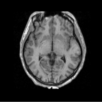
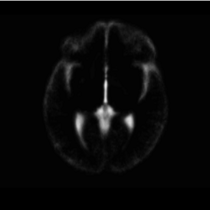
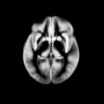
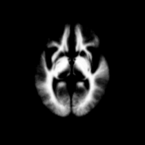
Atlas
Atlas was generated based on 82 scans and corresponding segmentations provided by Psychiatry Neuroimaging Laboratory, BWH. We registered the scans to a preselected template via Warfield et al. 2001.
Image Dimension = 256 x 256 x 124
Image Spacing = 0.9375 x 0.9375 x 1.5

|

|

|

|
| Template (T1) | CSF | GM | WM |
Result
Acknowledgment
The construction of the pipeline was supported by funding from NIH NCRR 2P41RR013218 Supplement.
Collaborators
Gabriella Szatmary, MD, PhD
Director of Neuro-Ophthalmology, Neuroimaging, Hattiesburg Clinic P.A., Mississippi
Tatjana Polgar, MBA
Research Assistant Neuro-Ophthalmology, Hattiesburg Clinic P.A., Mississippi
Citations
- Tustison NJ, Avants BB, Cook PA, Zheng Y, Egan A, Yushkevich PA, Gee JC N4ITK: Improved N3 Bias Correction, IEEE Trans Med Imag, 2010
- Pohl K, Bouix S, Nakamura M, Rohlfing T, McCarley R, Kikinis R, Grimson W, Shenton M, Wells W. A Hierarchical Algorithm for MR Brain Image Parcellation. IEEE Transactions on Medical Imaging. 2007 Sept;26(9):1201-1212.
- S. Warfield, J. Rexilius, P. Huppi, T. Inder, E. Miller, W. Wells, G. Zientara, F. Jolesz, and R. Kikinis, “A binary entropy measure to assess nonrigid registration algorithms,” in MICCAI, LNCS, pp. 266–274, Springer, October 2001.
- Johnson H.J., Harris G., Williams K. BRAINSFit: Mutual Information Registrations of Whole-Brain 3D Images, Using the Insight Toolkit, The Insight Journal, July 2007


