Difference between revisions of "EMSegmenter-Tasks:Template"
From Slicer Wiki
(→Result) |
|||
| (3 intermediate revisions by the same user not shown) | |||
| Line 20: | Line 20: | ||
-- how many set of images did you use <br> | -- how many set of images did you use <br> | ||
-- where did you get the data from <br> | -- where did you get the data from <br> | ||
| − | + | -- example images of atlas such as | |
| − | |||
| − | |||
| − | |||
| − | |||
| − | |||
{| class="wikitable" | {| class="wikitable" | ||
|- | |- | ||
| Line 40: | Line 35: | ||
=Result= | =Result= | ||
| + | show original scans with segmentation as in the example below <BR> | ||
[[Image:MRI-Human-Brain-T1.png|630px]]<br> | [[Image:MRI-Human-Brain-T1.png|630px]]<br> | ||
[[Image:MRI-Human-Brain-Labelmap.png|630px]] | [[Image:MRI-Human-Brain-Labelmap.png|630px]] | ||
Latest revision as of 21:53, 8 December 2010
Home < EMSegmenter-Tasks:TemplateReturn to EMSegmenter Task Overview Page
Contents
This wiki template can be used for new tasks
Description
-- Data Assumption
-- description of pipeline with citations to algorithms used
Anatomical Tree
-- Define Anatomical Tree used in mrml file - first list the complete name and then the abbreviating used in the mrml file
- root
- background (BG)
- air (AIR)
- background (BG)
Atlas
- how did you construct the atlas ?
-- what did you do for skull stripping or any other preprocessing
-- What did you do for group registration
-- how did you generate the segmentations
-- how many set of images did you use
-- where did you get the data from
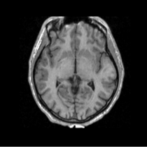
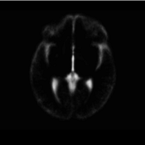
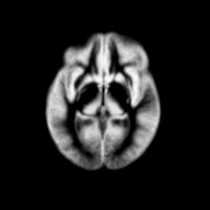
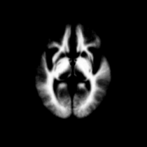
-- example images of atlas such as
|
|
|
|
| Template (T1) | CSF | GM | WM |
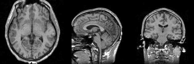
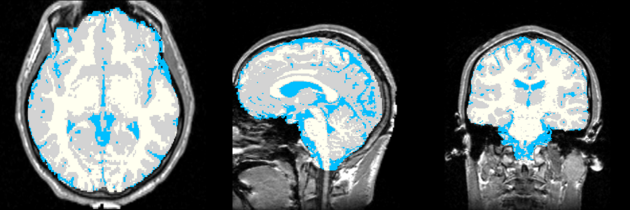
Result
show original scans with segmentation as in the example below
Acknowledgment
The construction of the pipeline was supported by funding from NIH NCRR 2P41RR013218 Supplement.
Citations
- Pohl K, Bouix S, Nakamura M, Rohlfing T, McCarley R, Kikinis R, Grimson W, Shenton M, Wells W. A Hierarchical Algorithm for MR Brain Image Parcellation. IEEE Transactions on Medical Imaging. 2007 Sept;26(9):1201-1212.