Difference between revisions of "User:Dpace"
From Slicer Wiki
m (Text replacement - "\[http:\/\/www\.slicer\.org\/slicerWiki\/images\/([^ ]+) ([^]]+)]" to "[http://www.slicer.org/w/img_auth.php/$1 $2]") |
|||
| (2 intermediate revisions by one other user not shown) | |||
| Line 10: | Line 10: | ||
|- | |- | ||
| style="background:#FFFFCC; color:black" valign="center"| Full head MRI scan | | style="background:#FFFFCC; color:black" valign="center"| Full head MRI scan | ||
| − | | id="MR-head" style="background:#FFFFCC; color:blue; font-size:110%" align="center"| '''[http://www.slicer.org/ | + | | id="MR-head" style="background:#FFFFCC; color:blue; font-size:110%" align="center"| '''[http://www.slicer.org/w/img_auth.php/4/43/MR-head.nrrd MR-head]''' |
| style="background:#FFFFCC; color:black" align="center"| [[Image:MR-head_screenshot.png |200px]] | | style="background:#FFFFCC; color:black" align="center"| [[Image:MR-head_screenshot.png |200px]] | ||
|- | |- | ||
| style="background:#FFFFCC; color:black" valign="center"| CT scan of chest | | style="background:#FFFFCC; color:black" valign="center"| CT scan of chest | ||
| − | | id="CT-chest" style="background:#FFFFCC; color:blue; font-size:110%" align="center"| '''[http://www.slicer.org/ | + | | id="CT-chest" style="background:#FFFFCC; color:blue; font-size:110%" align="center"| '''[http://www.slicer.org/w/img_auth.php/3/31/CT-chest.nrrd CT-chest]''' |
| − | | style="background:#FFFFCC; color:black" align="center"| [[Image: | + | | style="background:#FFFFCC; color:black" align="center"| [[Image:CT-chest_screenshot.png |200px]] |
|- | |- | ||
| style="background:#FFFFCC; color:black" valign="center"| A volume of tensors indicating brain diffusivity. Can be used with tractography. | | style="background:#FFFFCC; color:black" valign="center"| A volume of tensors indicating brain diffusivity. Can be used with tractography. | ||
| − | | id="DTI-head" style="background:#FFFFCC; color:blue; font-size:110%" align="center"| '''[http://www.slicer.org/ | + | | id="DTI-head" style="background:#FFFFCC; color:blue; font-size:110%" align="center"| '''[http://www.slicer.org/w/img_auth.php/0/01/DTI-Brain.nrrd DTI-head]''' |
| − | | style="background:#FFFFCC; color:black" align="center"| [[Image: | + | | style="background:#FFFFCC; color:black" align="center"| [[Image:DTI-Brain_screenshot.png |200px]] |
|- | |- | ||
| style="background:#FFFFCC; color:black" valign="center"| Registration example part 1: Head MRI of Meningioma at timepoint 1. | | style="background:#FFFFCC; color:black" valign="center"| Registration example part 1: Head MRI of Meningioma at timepoint 1. | ||
| − | | id="RegLib_C01_1" style="background:#FFFFCC; color:blue; font-size:110%" align="center"| '''[http://www.slicer.org/ | + | | id="RegLib_C01_1" style="background:#FFFFCC; color:blue; font-size:110%" align="center"| '''[http://www.slicer.org/w/img_auth.php/5/59/RegLib_C01_1.nrrd RegLib_C01_1]''' |
| style="background:#FFFFCC; color:black" align="center"| [[Image:RegLib_C01_1_screenshot.png |200px]] | | style="background:#FFFFCC; color:black" align="center"| [[Image:RegLib_C01_1_screenshot.png |200px]] | ||
|- | |- | ||
| style="background:#FFFFCC; color:black" valign="center"| Registration example part 2: Head MRI of Meningioma at timepoint 2. | | style="background:#FFFFCC; color:black" valign="center"| Registration example part 2: Head MRI of Meningioma at timepoint 2. | ||
| − | | id="RegLib_C01_2" style="background:#FFFFCC; color:blue; font-size:110%" align="center"| '''[http://www.slicer.org/ | + | | id="RegLib_C01_2" style="background:#FFFFCC; color:blue; font-size:110%" align="center"| '''[http://www.slicer.org/w/img_auth.php/e/e3/RegLib_C01_2.nrrd RegLib_C01_2]''' |
| style="background:#FFFFCC; color:black" align="center"| [[Image:RegLib_C01_2_screenshot.png |200px]] | | style="background:#FFFFCC; color:black" align="center"| [[Image:RegLib_C01_2_screenshot.png |200px]] | ||
|- | |- | ||
|} | |} | ||
Latest revision as of 12:39, 27 November 2019
This page contains sample data for getting started with 3D Slicer. These files can be downloaded from the File->Download Sample Data menu item.
Please don't delete these files from the wiki or add new ones.
| Description | Sample Data | Screenshot |
| Full head MRI scan | MR-head | 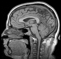
|
| CT scan of chest | CT-chest | 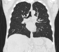
|
| A volume of tensors indicating brain diffusivity. Can be used with tractography. | DTI-head | 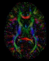
|
| Registration example part 1: Head MRI of Meningioma at timepoint 1. | RegLib_C01_1 | 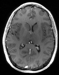
|
| Registration example part 2: Head MRI of Meningioma at timepoint 2. | RegLib_C01_2 | 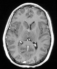
|