Difference between revisions of "Documentation/4.1/Training"
From Slicer Wiki
(Prepend documentation/versioncheck template. See http://na-mic.org/Mantis/view.php?id=2887) |
m (Text replacement - "http://wiki.slicer.org/slicerWiki/images/" to "http://www.slicer.org/w/img_auth.php/") |
||
| Line 149: | Line 149: | ||
{|width="100%" | {|width="100%" | ||
| | | | ||
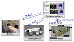
| − | *[http:// | + | *[http://www.slicer.org/w/img_auth.php/f/f1/OpenIGTLinkTutorial_Slicer4.1.0_JunichiTokuda_Apr2012.pdf Connecting IGT Device with OpenIGTLink] |
*Authors: Junichi Tokuda, BWH | *Authors: Junichi Tokuda, BWH | ||
|align="right"| | |align="right"| | ||
Latest revision as of 12:30, 27 November 2019
Home < Documentation < 4.1 < Training
|
For the latest Slicer documentation, visit the read-the-docs. |
Contents
Introduction: Slicer 4.1 Tutorials
- This page contains "How to" tutorials with matched sample data sets. They demonstrate how to use the 3D Slicer environment (version 4.1 release) to accomplish certain tasks.
- For tutorials for other versions of Slicer, please visit the Slicer training portal.
- For "reference manual" style documentation, please visit the Slicer 4.1 documentation page
- For questions related to the Slicer4 Compendium, please send an e-mail to Sonia Pujol, Ph.D
General Introduction
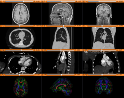
Slicer Welcome Tutorial
|
Slicer4Minute Tutorial
|
Slicer4 Data Loading and 3D Visualization
|
Tutorials for software developers
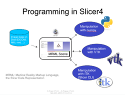
Slicer4 Programming Tutorial
|
Specific functions
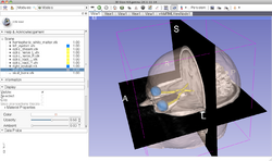
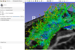
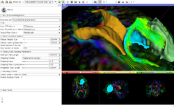
Slicer4 Diffusion Tensor Imaging Tutorial
|
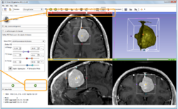
Slicer4 Neurosurgical Planning Tutorial
|
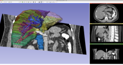
Slicer4 3D Visualization of DICOM images for Radiology Applications
|
Slicer4 Quantitative Imaging tutorial
|
Summer 2012 Tutorial contest (under construction)
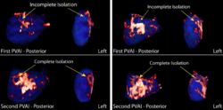
Automatic Left Atrial Scar Segmenter
|
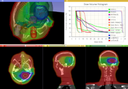
Qualitative and quantitative comparison of two RT dose distributions
|
Dose accumulation for adaptive radiation therapy
|
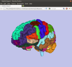
WebGL Export
|
OpenIGTLink
|
Additional resources
|
|
|
|
|
|
