Difference between revisions of "Documentation/Nightly/Modules/T1 Mapping"
Stevedaxiao (talk | contribs) |
Stevedaxiao (talk | contribs) |
||
| Line 42: | Line 42: | ||
<!-- ---------------------------- --> | <!-- ---------------------------- --> | ||
{{documentation/{{documentation/version}}/module-section|Panels and their use}} | {{documentation/{{documentation/version}}/module-section|Panels and their use}} | ||
| − | + | {| | |
| + | |[[Image:T1_Mapping_GUI.png|thumb|250px|T1 Mapping]] | ||
| + | | | ||
| + | * '''T1 Mapping Parameters''': | ||
<!-- | <!-- | ||
{{documentation/{{documentation/version}}/module-parametersdescription}} | {{documentation/{{documentation/version}}/module-parametersdescription}} | ||
Revision as of 02:42, 21 March 2015
Home < Documentation < Nightly < Modules < T1 Mapping
|
For the latest Slicer documentation, visit the read-the-docs. |
Introduction and Acknowledgements
|
Extension: T1_Mapping | |||||||
|
Module Description
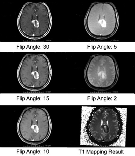
T1 mapping estimates effective tissue parameter maps (T1) from multi-spectral FLASH MRI scans with different flip angles.
Use Cases
- Takes an arbitrary number of multi-spectral FLASH images as input, and estimates the T1 values of the data for each voxel
- Read repetition time(TR), echo time(TE) and the flip angles from the Dicom header directly
- Prostate, brain, head & neck, cervix, breast and etc.
Tutorials
Panels and their use
Similar ModulesN/A ReferencesN/A Information for Developers
|


