Difference between revisions of "Main Page"
From Slicer Wiki
(→News) |
|||
| Line 9: | Line 9: | ||
<gallery widths="200px" perrow="4"> | <gallery widths="200px" perrow="4"> | ||
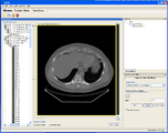
| − | Image: | + | Image:Gimias-running-slicer-module-2010-10-20.png | A set of slicer command line modules are now shipped as part of the [http://www.gimias.org Gimias] tool from UPF in Barcelona! |
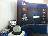
| − | + | Image:800px-NDI booth Slicer MICCAI2010.jpg | Northern Digital, makers of tracking systems widely used in image guided therapy, used Slicer and OpenIGTLink in their booth installation at [http://www.miccai2010.org/ MICCAI2010 in Beijing]. | |
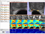
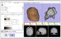
| + | Image:prostate_multiparametric_visualization.png | In this screenshot [[Modules:MainApplicationGUI-Documentation-3.6#Selecting_Among_Standard_Layouts|CompareView layout of 3D Slicer]] is used to facilitate visualization of multimodal MRI of the prostate | ||
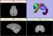
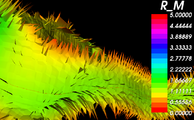
| + | Image:vervet_atlas_template.png| [http://www.nitrc.org/projects/vervet_atlas Vervet Probabilistic Atlas] constructed using 3D Slicer tools is available for download from NITRC. | ||
Image:Avf 3d voronoi big.png| A Voronoi diagram used for [[Modules:VMTKCenterlines|centerline computation]] of a vascular tree. | Image:Avf 3d voronoi big.png| A Voronoi diagram used for [[Modules:VMTKCenterlines|centerline computation]] of a vascular tree. | ||
| − | + | ||
</gallery> | </gallery> | ||
Revision as of 11:37, 22 October 2010
Contents
Welcome to the 3D Slicer Wiki pages!
Welcome to the Slicer Wiki Home Page.
How to use this site:
- The main slicer.org pages provide a guided tour to the application, training materials, and the development community. New users should start there because we try to keep it organized and up to date.
- These wiki pages are more free-form, with notes and reference pages that help support developers and users across multiple sites. You will find good information on the wiki, but it's possible that it's out of date or describes features that are still in the planning phase.
A set of slicer command line modules are now shipped as part of the Gimias tool from UPF in Barcelona!
Northern Digital, makers of tracking systems widely used in image guided therapy, used Slicer and OpenIGTLink in their booth installation at MICCAI2010 in Beijing.
In this screenshot CompareView layout of 3D Slicer is used to facilitate visualization of multimodal MRI of the prostate
Vervet Probabilistic Atlas constructed using 3D Slicer tools is available for download from NITRC.
A Voronoi diagram used for centerline computation of a vascular tree.
More images at the Slicer3 Visual Blog...
Slicer3 is:
- A software platform for the analysis and visualization of medical images and for research in image guided therapy.
- An algorithm and application development platform with a powerful plug-in architecture.
- A free, open source package available on multiple operating systems (Windows, MAC, Linux).
News
2010
- October
- Our friends and colleagues at UFP in Barcelona, Spain have released version 1.2 of the Gimias application. This new version includes an interface so that Gimias can discover and execute slicer command line modules to operate on data loaded in Gimias.
- September
- The first alpha versions of Slicer4 (the Qt version) are starting to become available. Contact the slicer-devel list if you want access to a binary installer for testing.
- A video has been nominated for official slicer bug fixing anthem ;)
- August
- The Slicer 3.6.1 patch release includes several bug fixes and other improvements. A 3.6.2 is in the works for sometime in the next few weeks to capture more bug fixes. See the list of fixed and outstanding issues for Slicer 3.6.x.
- July
- June
- May
- first (5/17) and second (5/25) release candidates for Slicer 3.6
- April
- Slicer factoids on Ohloh
- Feature Freeze month! Developers are working on bug fixes, documentation, and performance enhancement.
- Check out Andras Jakab's impressive surgery planning images
- March
- New infrastructure software is rapidly evolving in the context of the Common Toolkit working meetings.
- Planning underway for release 3.6 of 3D Slicer
- A slicer review on medfloss
- slicer's debian package page
- slicer ubuntu package that can be installed with the alpha version of Ubuntu 10.04
- A page with information about installing slicer3 on Debian has been started.
- We are starting to make plans for 3D Slicer version 4 (also known as Slicer4). A big step!
- January
2009
- December
- Lots of activity around the Port of Slicer to Qt
- New Annotation Tools are in the works
- November
- Seb Barre from Kitware has taken the lead in adding multiple cameras support to Slicer 3. The work is currently focused on multiple cameras and multiple views, but only one scene. The nightly build contains a new module, called cameras. Jim Miller has added a new layout, called Dual 3D Layout. .
- October
- Test of mobile friendly wiki skin
- September
- Serious work is beginning on a Slicer3 port to Qt.
- Attila Nagy and his colleagues created a very nice set of presentations and animations with slicer to illustrate 3D reconstruction as applied to otorhinolaryngology. (See Notes).
- August
- New images in the visual blog show integration of slicer and BioImageSuite.
- July
- Several bug fixes are being rolled into a 3.4.1 patch release, expected for beginning of August
- June
- NA-MIC Summer Project Week was a great success!
- New documentation is available, thanks to the Summer 2009 Tutorial Contest
- May
- Slicer 3.4 release
- The 3.4 release branch has been created in subversion
- A punch list of 3.4 bugs is being maintained on the Mantis Bug Tracker.
- March
- Development of slicer3.4 is still under way. To check the status, go to the bug tracker and search for the tag "3.4 Targeted fix". Our plan is to fix anything with that tag before branching the release. http://na-mic.org/Mantis/main_page.php The svn trunk for slicer3 is still in "feature freeze" mode, meaning that only fixes should be checked in. Any new development should occur in svn branches for now.
- Developers, please be sure to apply the "3.4 Targeted fix" to anything you are working on for the release.
- January
- A product release of Slicer 3.4 is scheduled for Feb/March
- There will be a code freeze on Feb 4, 2009
- Slicer3 and related research project were highlighted and demoed as part of the External Advisory Board meeting of the Neuroimage Analysis Center.
2008
- December
- Andrew Farke posted some excellent tutorials on the use of 3D Slicer in paleontology research.
- A training course on use of 3D Slicer by Radiologists was held at RSNA in Chicago.
- NCIGT is hosting a Project Week for developing Image Guided Therapy software that features many slicer projects.
- September
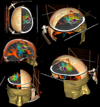
- Andras Jakab won second place in Kitware's medical image visualization contest!. The image combines slice viewing, models, volume rendering, and tractography applied to a gamma knife neurooncology radiotherapy planning case. Andras used slicer's registration tools to fuse CT and diffusion MR into a multimodal scene.
- A NA-MIC Workshop at MICCAI addressed the creation of ITK-based command line modules for 3D Slicer.
- Slicer user Attila Nagy presented on Slicer3 for inner ear imaging (Slides - Hungarian)
- Attila also provided patches to build Slicer3 and most of Slicer2 using the latest versions of Solaris and the optimized Sun C++ compilers.
- August
- July
- Airway Inspector is a powerful tool for lung image analysis build on slicer2. A port to slicer3 is getting under way in the next few weeks and is expected to take about 6 months.
- Slicer and the NA-MIC software environment were presented as part of the UCLA Institute for Pure and Applied Mathematics Summer School on Mathematics and Brain Imaging.
- A very productive working meeting between researchers at BWH and Yale resulted in successful connection of Slicer3 to the BrainLab commercial image guided neurosurgery system.
- June
- May
- Slicer 3.2 release
- Slicer3 included in review paper on "Rapid Development of Medical Imaging Tools with Open Source Libraries"
- April
- March
- A new 3D Slicer Wikipedia entry has been created.
- February
- February 20th, 2008, Spring 2008 Slicer3 User Training Workshop in the SPL at 1249 Boylston St. Boston.
- January
- Wishlist for tutorials
- Lots of Slicer development activity as part of NA-MIC All Hands Meeting and Winter Project Week
2007
- December
- A three-day retreat for Slicer in Image Guided Therapy was held in Boston.
- November
- New builds are now available for Linux 64, Linux 32, Windows, Darwin PPC, and Darwin x86
- June
- Lots of Slicer3 work at the Project Weeks
- March
- New Image Guided Therapy (IGT) functionality for tracking surgical instruments added by developers working at the National Center for IGT.
- February
- February 7-8, 2007. Slicer3 Mini-Retreat, Boston
- Resulted in the implementation of a Python interface to Slicer3.
- January
- Slicer3 developers are getting together in Salt Lake City for a "programming half-week" as part of the NA-MIC All-Hands Meeting
- Lots of new developer documentation added!
- Slicer3 beta released
- New bug tracker installed
2006
- December
- There was a big meltdown of the na-mic.org server in December, and some things were lost including wiki history, slicer3 bug tracker and doxygen, and some of the older download files. Please bear with us as we pick ourselves up and carry on.
- November
- Slicer3 Alpha builds available for Linux (32 and 64 bit) and windows!
- October
- Slicer3 development version shown at the BIRN All Hands Meeting as the platform for the BIRN Query Atlas Project and overall visualization issue in BIRN. See these slides for more information.
- September
- July
- There was active development of Slicer3 at and around the NA-MIC Programming/Project week.
- New Slicer3 wiki page organization is under way. Please send feedback to the slicer-developers mailing list.
For Wiki Administration
- Create user account
- Users List
- Authorized users can commit edited web pages through slicer's wiki2web interface
